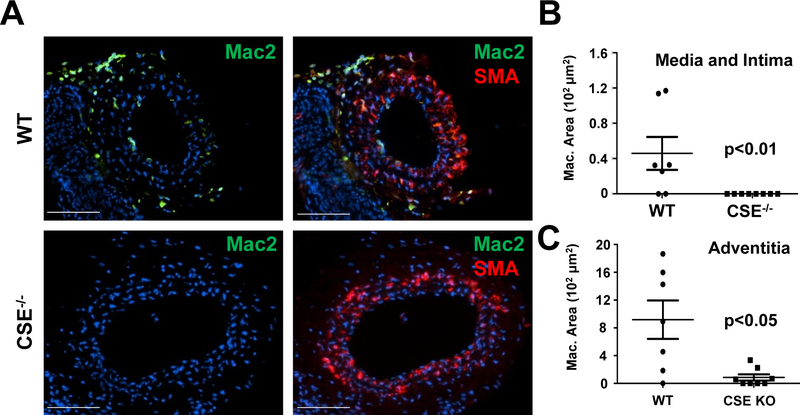
Figure 3. Absence of macrophages in ligated CSE−/− carotids.
(A) 7 days after partial carotid ligation in WT and CSE−/− mice, mouse carotid arteries were immunostained for macrophages (Mac-2) and smooth muscle actin (SMA). (B/C) Macrophages in (B) tunica intima, media, and (C) adventitia were quantified by Mac-2 positive area (scale bar = 100 μm, n=7–8, p<0.05 by (B) Mann-Whitney U Test and (C) Students T-test).

