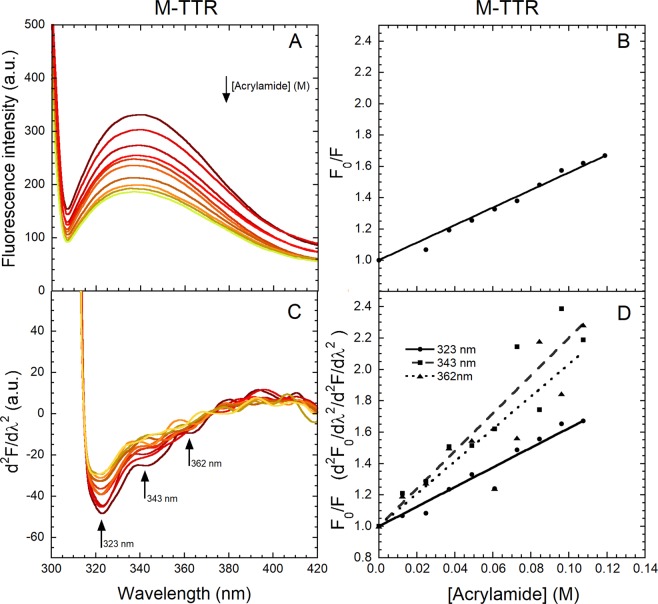
Figure 3.
Quenching of M-TTR fluorescence spectra by acrylamide. (A) Fluorescence spectra (excitation 290 nm, slits 2.5 and 18 nm for ex and em, respectively) of 2.2 µM M-TTR following progressive additions of small volumes of 2.5 M acrylamide in 20 mM phosphate buffer, pH 7.4, 25 °C. (B) Plot of the ratio of total fluorescence intensity in the absence (F0) and presence (F) of acrylamide (F0/F) versus acrylamide concentration. The straight line represents the best fit of the data points to the Stern-Volmer linear function (Eq. 1) to determine the KSV value. (C) Corresponding second derivative spectra with arrows indicating the major peaks at 323, 343 and 362 nm. (D) F0/F versus acrylamide concentration for the three indicated second derivative peaks, where F0 and F values are second derivative values. The lines through the data represents the best fit to the Stern-Volmer linear function (Eq. 1).

