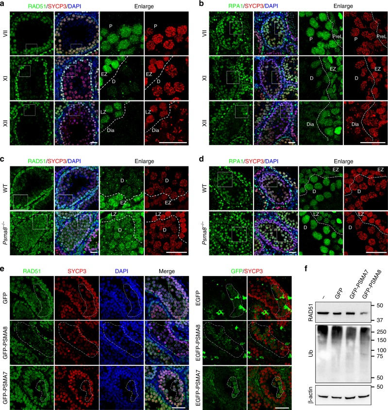
Fig. 4.
Failure in RAD51 and RPA1 degradation in PSMA8-deleted spermatocytes. a, b Immunostaining of RAD51 (a) and RPA1 (b) in PD25 testes sections. SYCP3 (red) and DAPI (blue) staining showing the stages of spermatogenesis. The different stages of seminiferous tubules are indicated by roman numerals. The regions bordered with a dashed box are enlarged on the right two panels. Scale bars, 25 μm. EZ early zygonema, Dia diakinesis spermatocytes. c–d Immunostaining of RAD51 (c) and RPA1 (d) in testes sections derived from wild-type and Psma8−/− males at the age of PD25. Scale bars, 25 μm. e Immunostaining of RAD51 in sections of testes electroporated with plasmids encoding green fluorescent protein (GFP), GFP-PSMA8, and GFP-PSMA7, respectively. Immunostaining of GFP was shown on the right to indicate the GFP-expressing cells, which were bordered with dashed circles. Scale bars, 50 μm. f Western blotting showing the levels of RAD51 and ubiquitination in testes electroporated with GFP, GFP-PSMA7, or GFP-PSMA8

