Fig. 5.
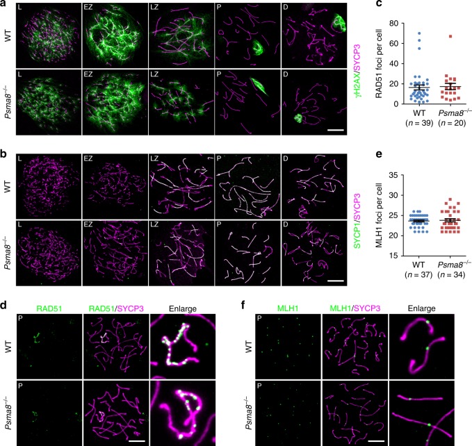
Meiotic recombination is less affected by PSMA8 deletion. a, b Staining of γH2AX (a) and SYCP1 (b) in nuclear surface spreads derived from wild-type (WT) and Psma8−/− males at PD42. Scale bars, 10 μm. c, d Immunostaining of RAD51 (d) in the nuclear surface spreads derived from WT and Psma8−/− males at PD42 and the quantification of RAD51 foci (c) in WT and Psma8−/− spermatocytes at pachytene stage. Scale bar, 10 μm. n = 39 for WT spermatocytes and n = 20 for Psma8−/− spermatocytes. Median focus numbers are marked. Error bars indicate S.E.M. e, f Immunostaining of MLH1 (f) in nuclear surface spreads derived from WT and Psma8−/− males at PD42. The quantification of MLH1 foci in WT and Psma8−/− spermatocytes at the pachytene stage is shown in e. Scale bar, 10 μm. n = 37 for WT spermatocytes and n = 34 for Psma8−/− spermatocytes. Median focus numbers are marked. Error bars indicate S.E.M.

