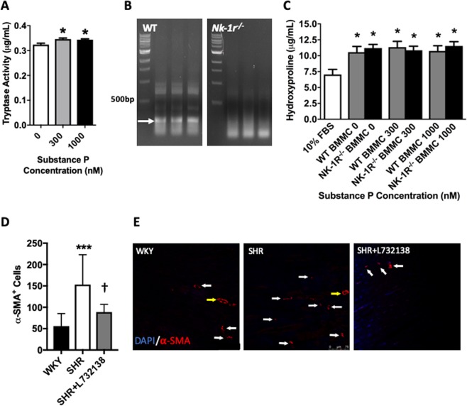
Figure 3.
SP does not cause MCs to produce sufficient amounts of pro-fibrotic mediators to influence fibroblast function in vitro, but does increase myofibroblast number in vivo. (A) Tryptase activity in the media of BMMCs treated with SP (n = 4); (B) PCR showing disruption of the PCR product for the NK-R in BMMCs derived from Nk-1r−/− mice; (C) Hydroxyproline production by 3T3 fibroblasts in response to conditioned media from SP treated BMMCs (n = 15). SP concentrations refer to the treatment of the BMMCs. 10% FBS served as a control. All values are mean ± SEM; *P < 0.05 vs SP 0 nM for (A) and vs 10% FBS for (C). (D) Quantification of α-smooth muscle actin (SMA)+ myofibroblasts in LV sections from WKY, SHR, and SHR treated with L732138 (n = 8/group). Values are mean ± SD; ***P < 0.001 vs WKY, †p < 0.05 vs SHR; (E) Representative images of α-SMA+ myofibroblasts (red = α-SMA). Blue = DAPI nuclear labeling, yellow arrow = blood vessels, white arrow = myofibroblast.

