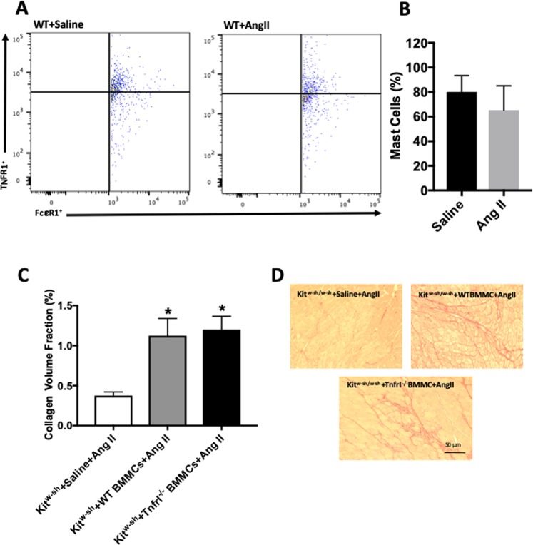
Figure 5.
MC-specific TNFRI does not play a functional role in the activation of MCs and cardiac fibrosis in vivo. (A) Representative flow cytometry scatter plots indicating the percentage of MCs that possess TNFRI, and (B) quantitative flow cytometry analysis of the percentage of MCs possessing TNFRI in left ventricles from wild type mice receiving saline or angiotensin II; (C) Collagen volume fraction for Kitw-sh/w-sh mice receiving angiotensin II with no BMMCs (n = 7), Kitw-sh/w-sh mice receiving wild type BMMCs and angiotensin II (n = 7), and Kitw-sh/w-sh receiving TnfrI−/− BMMCs and angiotensin II (n = 8); and (D) corresponding representative picrosirius red images. All values are mean ± SEM. *P < 0.05 vs Kitw-sh/w-sh mice receiving angiotensin II with no BMMCs.

