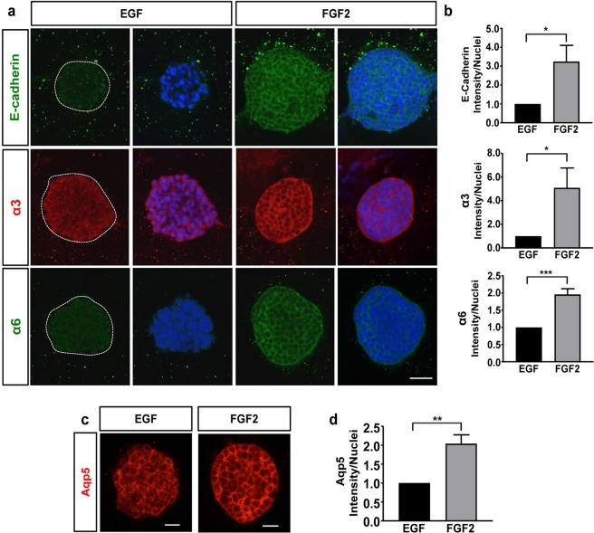
Figure 4.
FGF2 promotes the expression of E-cadherin, laminin-binding integrins, & Aqp5. Representative confocal images of mSG-PAC1 spheroids cultured in EGF or FGF2-containing medium for five days in a Matrigel/collagen I matrix and stained for (a) E-cadherin, integrin α3 subunit, or integrin α6 subunit and DNA, or (c) Aqp5. Images are maximum projection images of five z-slices acquired at 40X taken in 0.4 μm steps. (b & d) Fluorescence intensity was normalized to the number of nuclei per field from 13 spheres from three independent experiments and plotted relative to the expression in EGF ± s.e.m. Size bar, 50 μm. Data was analyzed by Student’s T-test. P < 0.05–0.0001, as indicated.

