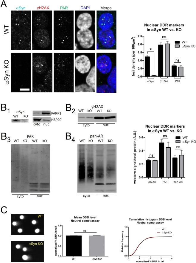
Figure 2.
Alpha-synuclein knock-out does not alter DSB levels at baseline in HAP1 cells. (A) SNCA knock-out (αSyn KO) does not alter baseline levels of nuclear γH2AX or PAR foci (foci density per 100 μm2): WT αSyn = 1.23 ± 0.10, αSyn KO αSyn = 0.06 ± 0.02, unpaired t-test p < 0.0001; WT γH2AX = 1.96 ± 0.14, αSyn KO αSyn = 2.03 ± 0.15, unpaired t-test p = 0.7422; WT PAR = 0.67 ± 0.07, αSyn KO PAR = 0.60 ± 0.09, unpaired t-test p = 0.5452; WT N = 326 nuclei, αSyn KO N = 252 nuclei). Scale bar 5 μm. (B) Left: (B1) Western blotting shows expected absence of αSyn protein in αSyn KO cells. Subcellular fractionation was used to purify nuclear and cytoplasmic proteins, as demonstrated by relative enrichment of the nuclear protein PARP1 in the nuclear fraction and the cytosolic protein HSP90 in the cytoplasmic fraction. Using this approach, blotting for nuclear γH2AX (B2), PAR (B3) and pan-(mono- & poly-) ADP-ribose (B4) showed no significant difference between WT and αSyn KO cells at baseline, when normalized to total protein levels (using REVERT stain, not shown). Right: Group data: WT γH2AX = 0.25 ± 0.01, αSyn KO γH2AX = 0.26 ± 0.02, unpaired t-test p = 0.9682; WT PAR = 0.52 ± 0.05, αSyn KO PAR = 0.45 ± 0.08, unpaired t-test p = 0.4883; WT pan-AR = 0.29 ± 0.04, αSyn KO pan-AR = 0.34 ± 0.07, unpaired t-test p = 0.6365; WT N = 3, αSyn KO N = 3 biological replicates). (C) Neutral comet assay shows no difference in levels of DSBs between WT and αSyn KO cells at baseline. Left: Comet images, middle: group data (normalized % DNA in tail: WT = 1.00 ± 0.01, αSyn KO = 1.00 ± 0.01, unpaired t-test p = 0.8860; WT N = 3016, αSyn KO N = 4171), right: cumulative probability histogram showing superimposable distributions.

