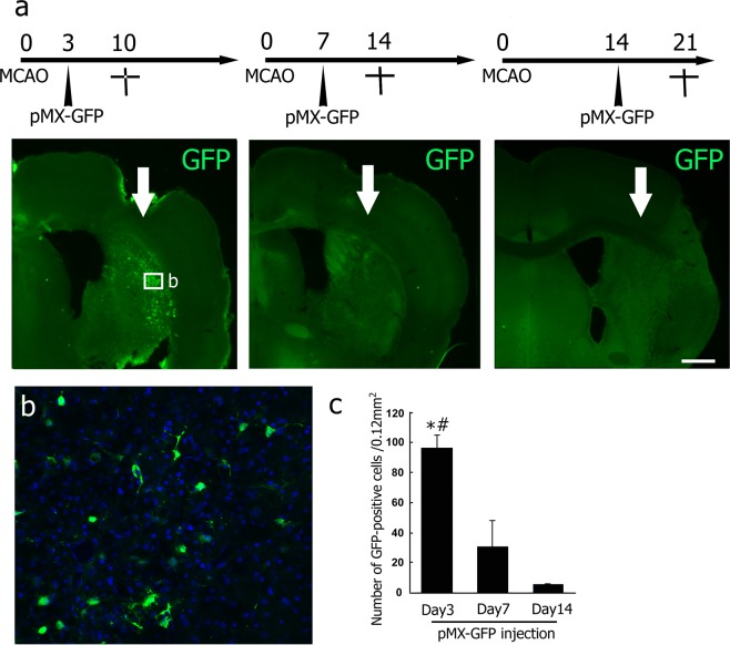
Figure 1.
(a) Time point-dependent efficacy of viral infection at 3, 7, and 14 d of injection after tMCAO (arrows; site of injection). (b) High-power confocal images of the infected area indicated by the boxed areas in A. Some Hoechst 33258-positive cells (blue) expressed detectable GFP (green). (c) Number of GFP-positive cells in a post-stroke brain section. Values are means ± S.D. *p < 0.05 vs Day 7, #p < 0.05 vs Day 14. Scale bar in (a) 200 μm, and in (b) 50 μm.

