
Fig. 16.
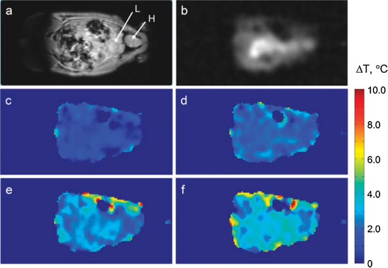
In-vivo thermometry in rat using TmDORMA‒. Representative coronal slices from 3D 1H MRI of water (a). Heart and liver labeled H and L, respectively. Methyl signal from TmDOTMA− (b) and temperature change maps computed from the phase shift in TmDOTMA− images as the rectal temperature changed from 35.7 to 36.4 (c), 37.1 (d), 37.6 (e) and 38.1 (f) °C. The TmDOTMA− dose was 0.61 mmol/kg body weight and the total imaging time was ~3 min. From ‘‘Non-invasive temperature imaging with thulium 1,4,7, 10-tetraazacyclododecane-1,4,7,10-tetramethyl-1,4,7, 10-tetraacetic acid (TmDOTMA-)’’ Sait Kubilay Pakin, S. K. Hekmatyar, Paige Hopewell, Andriy Babsky and Navin Bansal. NMR Biomed. 2006;19:116–124. Fig. 4. Reprinted with permission.