Figure 2.
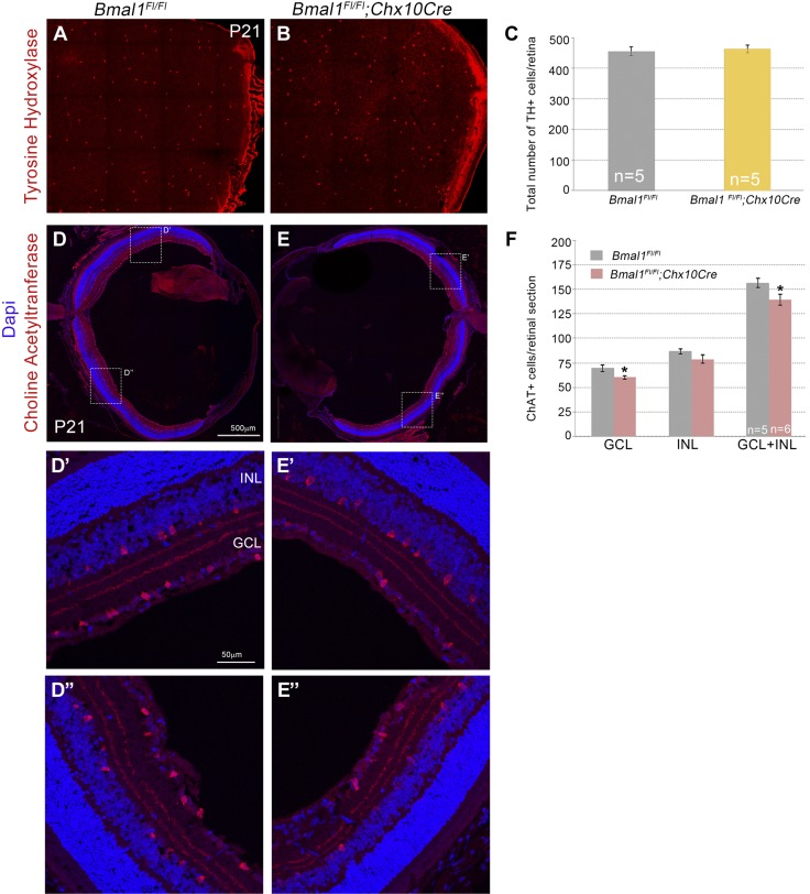
Loss of Bmal1 results in a decrease in cholinergic amacrine cells but not dopaminergic amacrine cells. A, B) Representative images of TH+ dopaminergic amacrine cells. C) Densities of TH+ cells were unaltered between control and Bmal1 CKO retinas. D, E) Representative images of ChAT+ cholinergic amacrine cells. D′, D″) Higher magnification images of the boxed area in D. E′, E″) Higher magnification images of the boxed area in E. F) Numbers of ChAT+ cells were significantly reduced in the Bmal1 CKO retinas compared with the control, and this reduction was more prominent in the ganglion cell layer. Scale bar, 500 µm (D, E), 50 µm (D′–E″). Error bars ± sem, n = 5–6. *P < 0.05.

