Figure 4.
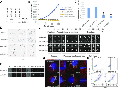
Constitutive knockdown of NCAPG significantly impairs HCC cell survival. A) Western blot showing the successful knockdown of NCAPG in HCCLM3 cells transduced with lentivirus expressing shRNAs targeting NCAPG (shNCAPG-1 and -2) compared with either control shRNAs (shControl-1 and -2). with GAPDH as endogenous control. B) Relative cell growth showing cells constitutively expressing shRNAs targeting NCAPG (shNCAPG-1 and -2) failed to proliferate in vitro. C) Mean number of cell divisions per starting cell during 24-h period (recorded using time-lapse live-cell imaging) showed significantly fewer cell divisions in constitutive NCAPG knockdown cells compared with control cells. D) Representative images showing cells with constitutive knockdown of NCAPG died after cell division (red arrows). Magnification value, ×10. E) Time-lapse confocal microscopy on H2B–enhanced green fluorescent protein-expressing HCCLM3 cells showing slowed kinetics in condensing and decondensing chromosomes in mitosis in constitutive NCAPG knockdown cells compared with control cells. Magnificaiton value, ×40. F) Immunofluorescence staining showed the cellular localization of DNA, NCAPG, α-tubulin, and γ-tubulin in mitosis. Constitutive NCAPG knockdown cells showed normal centrosome assembly (red arrows) and furrowing during cytokinesis (blue arrows) compared with control cells. Magnification value, ×60 G) Superresolution confocal microscopy images showing extensive fragmentation of mitochondrial network (region 5–8) in cells with constitutive knockdown of NCAPG compared with control cells (region 1–4), revealed through staining using MitoTracker (red) and Hoechst (blue) (Sanofi, Paris, France). Magnification value, ×120. Scale bars, 10 µm. H) FACS analysis showed significantly higher cell deaths in constitutive NCAPG knockdown cells, as indicated by 7-aminoactinomycin D (7-AAD)–positive cells. DIC, differential interference contrast. *P < 0.05, **P < 0.01.

