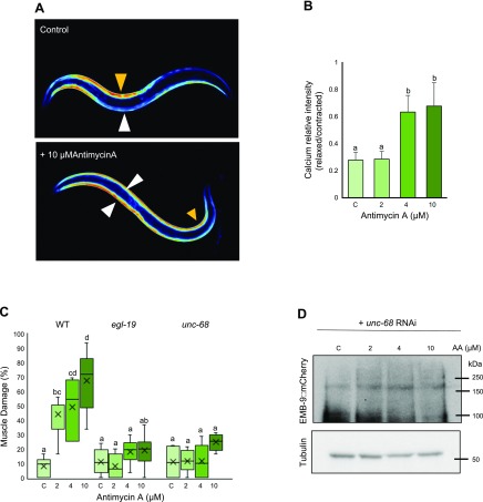
Figure 4.
Inhibition of calcium overload could rescue muscle damage and EMB-9 degradation in Antimycin A–treated C. elegans. A, B) Animals expressing the calcium sensor GCaMP3.35 were age synchronized at the L1 stage and grown to young adulthood at 20°C. Adults were treated with 0 (C, control), 2, 4, and 10 µM Antimycin A for 36 h, after which they were imaged using fluorescence microscopy with objective lens, ×10 magnification (UPlanAPO, Olympus) (A) and the calcium level quantified (B) using ImageJ software. A significant rise in intracellular calcium was observed following treatment with 4 and 10 µM Antimycin A. Five animals were analyzed for each condition. C) Synchronized adult animals [WT, unc-68(r1162) V and egl-19(ad695) IV] were treated with 0 (C, control), 2, 4, and 10 µM Antimycin A for 36 h and scored for muscle damage as performed in Fig. 1B. When compared with WT animals, there was significant rescue or decrease in muscle damage in both mutants at 2, 4, and 10 µM Antimycin A. Number of muscle images/conditions (167, 218, 301, 273, 64, 80, 74, and 42 from left to right). D) Synchronized adult worms expressing emb-9::mCherry were treated with 0 (C, control), 2, 4, and 10 µM Antimycin A along with unc-68 double-stranded RNA–containing bacteria. After 36-h treatment with Antimycin A, animals were subjected to lysis, and Western blot was performed to analyze the amount of EMB-9 protein using anti-mCherry antibody. No significant decrease in EMB-9 was observed with unc-68 RNAi after Antimycin A treatment (3 biologic repeats). Letters (a–d) on top of bars indicate statistical significance; X, median value.

