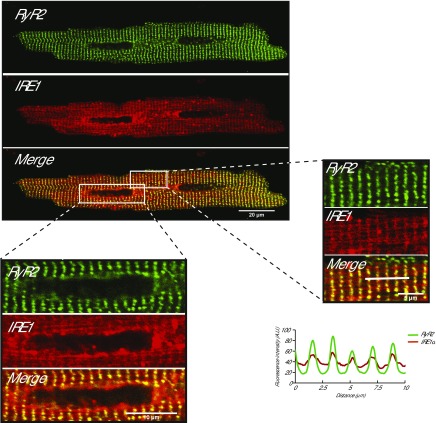
Figure 2.
IRE1α in isolated cardiomyocytes. Isolated rat cardiomyocytes from GFP-tagged RyR2 knock-in transgenic mice (35) were transduced with adenovirus packed with the RFP-tagged IRE1α (IRE1α). Large magnification of the sarcomere and perinuclear areas is shown as indicated by the boxes. Graphic representation of overlap between RyR2 and IRE1α is shown. The white bars indicate the scanned area represented in the graphs.

