Figure 3.
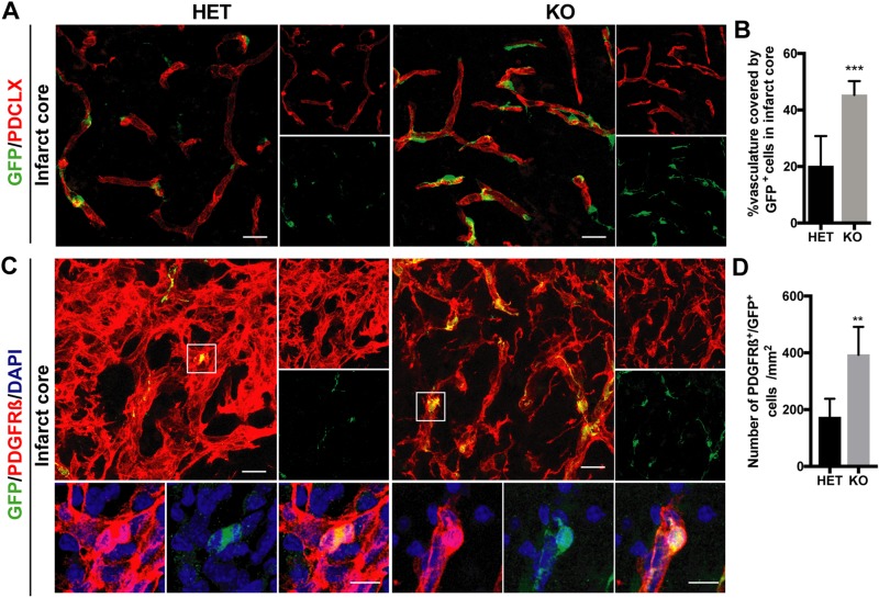
RGS5-KO mice have higher pericyte coverage 7 d after stroke. A) Representative confocal pictures of the infarct core of GFP+ pericytes (green) and the vasculature [podocalyxin (PDCLX), red]. Right panels show single channels of PDCLX and GFP, respectively. B) Quantification GFP+ pericyte coverage of the vasculature in the infarct core. C) Representative confocal pictures of the infarct core of PDGFR-β (red) and GFP (green) staining. Boxes indicate where higher-magnification pictures were taken. Right panels show single channels of PDGFR-β and GFP staining, respectively. Lower panels show higher magnification of pericytes colabeling for PDGFR-β and GFP. D) Quantification of the numbers of PDGFR-β+ and GFP+ pericytes in RGS5-HET and RGS5-KO mice in the infarct core. n = 6; data are represented as means ± sd. Scale bars, 20 μm (10 μm in higher magnification). **P < 0.01, ***P < 0.001, Student’s t test.

