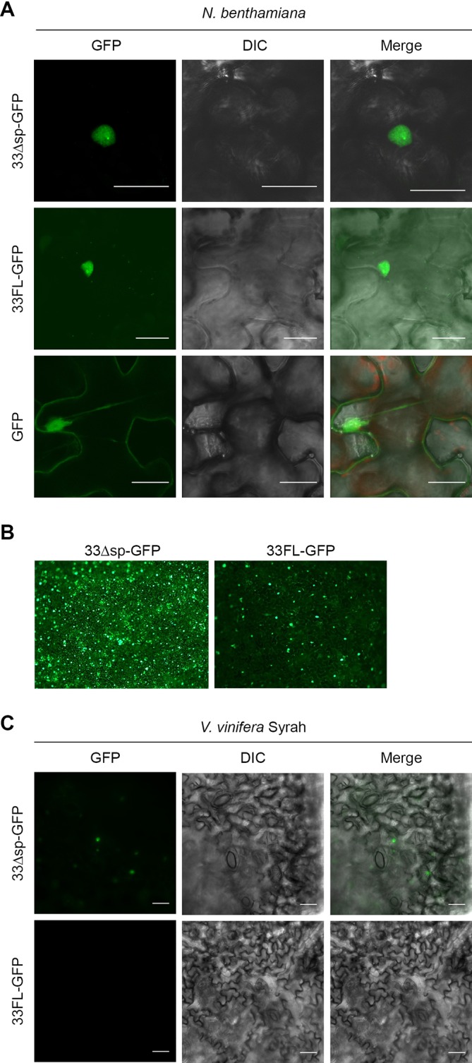
Fig 7. Pv33 localizes to the nucleus.

(A) Confocal microscopy images following infiltration of N. benthamiana leaves with Agrobacterium carrying GFP translational fusions of Pv33 either lacking (33Δsp-GFP) or including (33FL-GFP) the signal peptide. GFP alone was used as control. (B) Low magnification epifluorescence microscopy images of 33ΔSP-GFP and 33FL-GFP infiltrations. (C) Epifluorescence microscopy images following infiltration of V. vinifera cv Syrah leaf discs with Agrobacterium carrying 33ΔSP-GFP and 33FL-GFP. Pictures were taken 2 days after agroinfiltration. Experiments were repeated 5 times for N. benthamiana and twice for V. vinifera, with the same results. Bar shows 25 µm.
