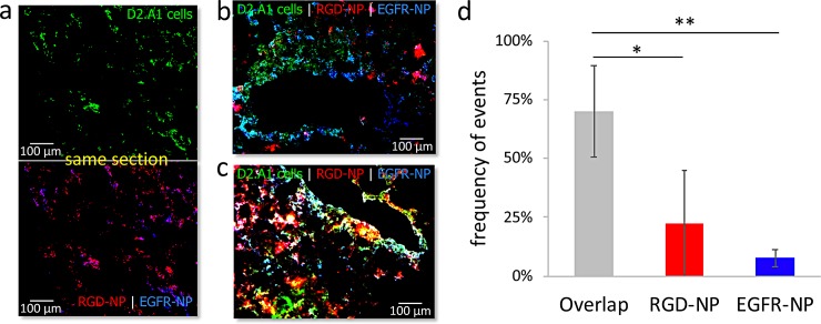
Fig 5. Histological evaluation of the microdistribution of RGD-NP and EGFR-NP nanoparticles in metastasis in the lungs of mice.
(a) Representative fluorescence image of lung tissue shows dispersion D2.A1 metastatic cancer cells (top; 20X magnification). After 3 h from injection of a cocktail containing Alexa 350-labeled EGFR-NP and Alexa 568-labeled RGD-NP, the two targeting variants colocalized in locations with metastatic cancer cells (bottom). Different regions with metastatic cancer cells were predominantly targeted by (b) EGFR-NP or (c) RGD-NP (green: D2.A1 cancer cells; red: RGD-NP; blue: EGFR-NP). (d) A pixel-by-pixel quantification indicates individual events for EGFR and RGD-NP or their overlap (n = 3, grouped analysis ANOVA; correct for multiple comparisons using the Holm−Sidak method. P values: *0.024, **0.002).

