. 2019 Jun 26;8:e45418. doi: 10.7554/eLife.45418

Figure 4. Craniofacial skeleton defects in Rbfox2 mutant embryos.

Alizarin red and Alcian blue stainings for ossified and chondrified tissues, respectively, of control (A–B), (D–E), (G), and I–J) and Rbfox2Pax3-CKO (C), (F), (H), and (K) skeleton at E17.5 (n = 8 controls, n = 6 Rbfox2Pax3-CKO). Alizarin red and Alcian blue stainings of control (L), (N), (P), and (R) and Rbfox2Wnt1-CKO (M, O, Q and S) skeleton at E18.5 (n = 8 controls, n = 5 Rbfox2Wnt1-CKO). Neural crest and mesoderm contribution to craniofacial bones are represented in green and pink color respectively (A, D and I). Dorsal (A–C and L–M), lateral (D–F and N–O) and ventral (I–K and R–S) view of skulls. Both Rbfox2Pax3-CKO (C), (F), (H), and K) and Rbfox2Wnt1-CKO (M), (O), (Q), and S) embryos demonstrate severe hypoplasia and diminished ossification of many neural crest-derived bones. Asterisks (*) represent the missing PPPL bone in Rbfox2 mutant embryos (K and S). AS, alisphenoid; BO, basioccipital; BS, basisphenoid; EO, exoccipital; F, frontal bone; IP, interparietal; MD, mandible; MX, maxilla; N, nasal; NC, nasal capsule; P, parietal bone; PL, palatine; PMX, premaxilla; PPMX, palatal process of maxilla; PPPL, palatal process of palatine; PPPMX, palatal process of premaxilla; PT, petrous part of temporal bone; SO, supraoccipital; SQ, squamous.

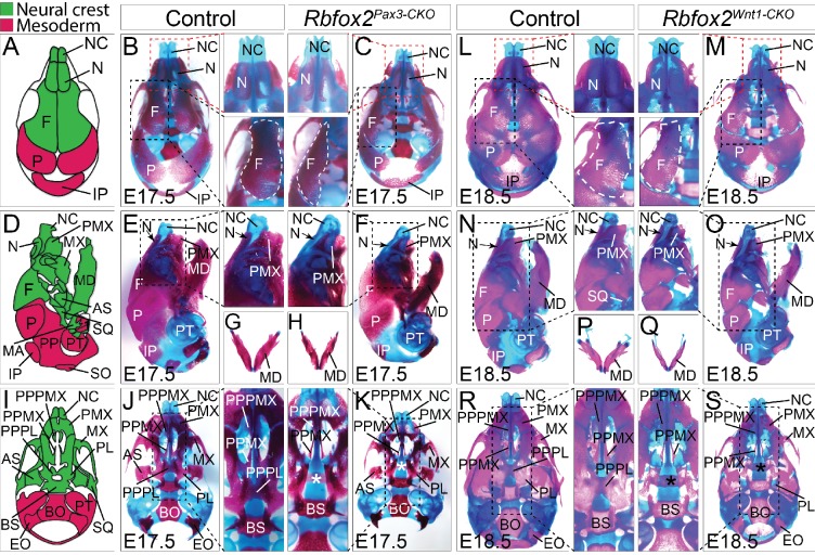
Figure 4.

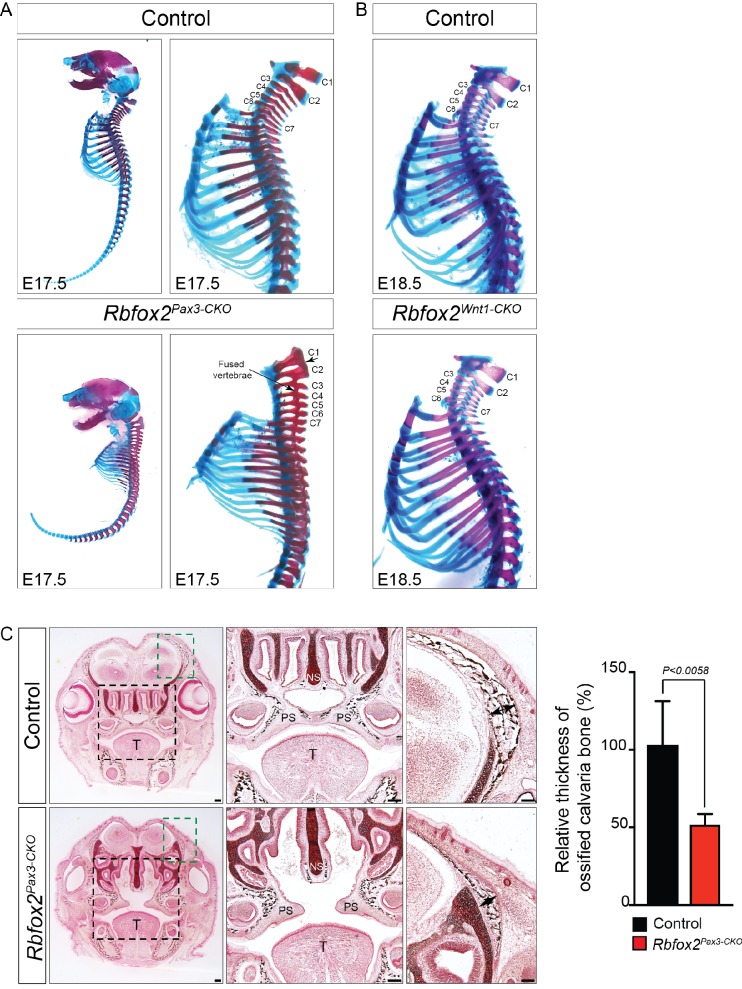
Figure 4—figure supplement 1. Skeletal defects in Rbfox2 mutant embryos.

Figure 4—figure supplement 1.

Alizarin red and Alcian blue stainings for ossified and chondrified tissues, respectively, of control and Rbfox2Pax3-CKO skeleton at E17.5 (n = 8 controls, n = 6 Rbfox2Pax3-CKO). Lateral view of full skeletons and higher magnification of cervical and thoracic parts of the vertebral column (A). Cervical and thoracic parts of control and Rbfox2Wnt1-CKO skeleton at E18.5 (n = 8 controls, n = 6 Rbfox2Wnt1-CKO) (B). Transverse sections through the palatal regions of E17.5 control and Rbfox2Pax3-CKO embryos are stained for mineralized bone (black) by Von Kossa staining (C). Black staining represents bone and red staining represents connective tissues (n = 4 controls, n = 4 Rbfox2Pax3-CKO). In comparison to controls, there was a reduced thickness of ossified calvaria bone in the Rbfox2Pax3-CKO embryos (double-headed black arrow). Scale bars are 100 μm respectively.
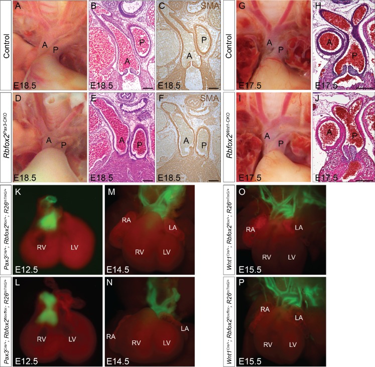
Figure 4—figure supplement 2. Neural crest contribution to the cardiac OFT is not affected in Rbfox2 knockout embryos.

Figure 4—figure supplement 2.

Gross morphology and histological analyses show no change in the patterning of the great vessels between controls (A–C and G–H) and Rbfox2Pax3-CKO (D–F); n = 10 controls, n = 10 Rbfox2Pax3-CKO) or Rbfox2Wnt1-CKO (I–J); n = 7 controls, n = 5 Rbfox2Wnt1-CKO) mouse embryos. Images of aortic arches in E18.5 control (A) and Rbfox2Pax3-CKO (D) embryos. H and E staining (B and E) and SMA immunohistochemistry (C and F) on sections through aortic arches from control (B–C) and Rbfox2Pax3-CKO (E–F) embryos. Images of aortic arches in E17.5 control (G) and Rbfox2Wnt1-CKO (I) embryos. H and E staining on sections through aortic arches from control (H) and Rbfox2Wnt1-CKO (J) embryos. Hearts from E12.5 and E14.5 Pax3Cre/+;Rbfox2flox/+;R26mTmG/+ (K and M; n = 4) and Pax3Cre/+;Rbfox2flox/flox;R26mTmG/+ (L and N; n = 5) embryos show labeled Pax3-derived neural crest derivatives (green) in the outflow tract. Heart from E15.5 Wnt1Cre/+;Rbfox2flox/+;R26mTmG/+ (O; n = 5) and Wnt1Cre/+;Rbfox2flox/flox;R26mTmG/+ (P); n = 5) embryos show labeled Wnt1-derived neural crest derivatives (green) in the outflow tract. A, Aorta; P, Pulmonary trunk; RA, right atrium; LA, left atrium; RV, right ventricle; LV, left ventricle. Scale bars are 200 μm respectively.
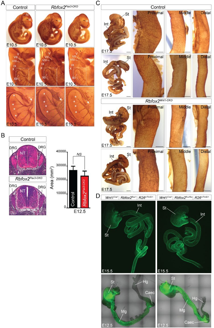
Figure 4—figure supplement 3. Development of peripheral and enteric nervous system in Rbfox2 mutant embryos.

Figure 4—figure supplement 3.

Whole mount neurofilament (2H3) immunostaining of one E10.5 control and two Rbfox2Pax3-CKO embryos are presented (A). White asterisks indicate nerve roots exiting the ventral neural tube. Cranial nerves are referred to as oculomotor (III), trochlear (IV), trigeminal (V), facial/vestibulocochlear (VII/VIII), glossopharyngeal (IX), vagal (X) and hypoglossal (XII). Oculomotor (III) and trochlear (IV) nerves are deformed (black arrow). The hypoglossal nerve (XII) is disorganized and sometime shorter (white arrows) in Rbfox2Pax3-CKO embryos (n = 6 controls, n = 5 Rbfox2Pax3-CKO) (A). Quantitation of dorsal root ganglia in control and Rbfox2Pax3-CKO embryos (n = 4 controls, n = 4 Rbfox2Pax3-CKO) (B). Whole-mount immunostaining of the gastrointestinal tract at E17.5 in two controls and two Rbfox2Wnt1-CKO embryos with the anti-neurofilament antibody 2H3 are presented (C). Scale bars are 500 μm respectively. Higher magnification of the proximal, middle and distal part of the intestine is presented (n = 4 controls, n = 4 Rbfox2Wnt1-CKO). Scale bars are 200 μm respectively. Enteric neural crest lineage tracing in E12.5 and E15.5 control (Wnt1Cre/+:Rbfox2flox/+:R26mTmG/+, n = 3) and Rbfox2 mutant (Wnt1Cre/+:Rbfox2flox/flox:R26mTmG/+, n = 3) embryos (D). DRG, dorsal root ganglion; NT, neural tube; St, stomach; Int, intestine; Mg, midgut; Hg, hindgut; Caec, Caecum.
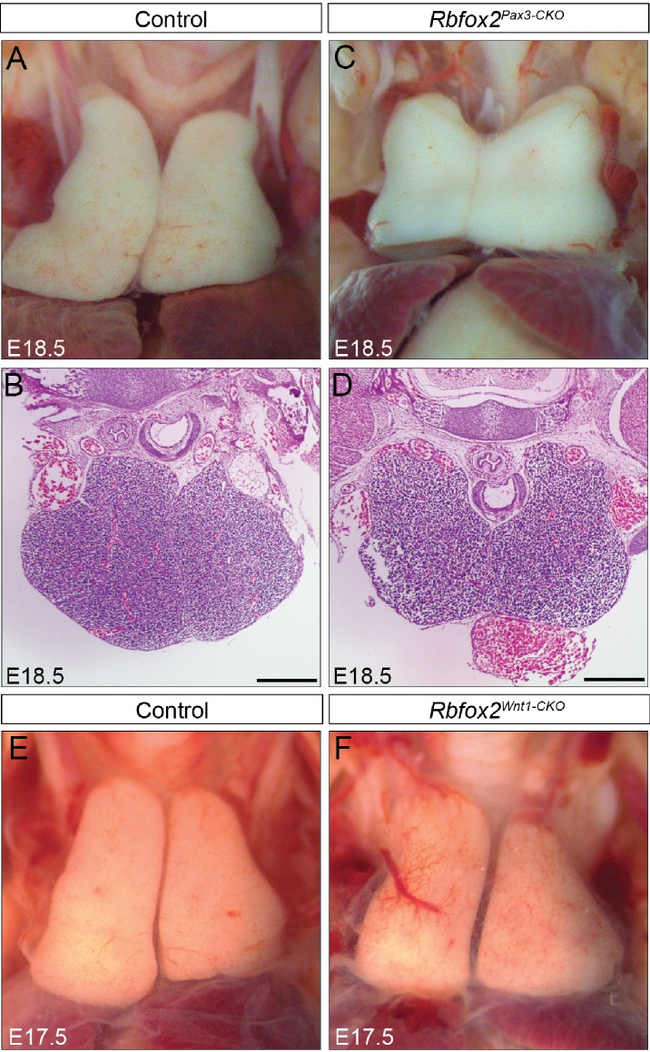
Figure 4—figure supplement 4. Thymus development is grossly intact in Rbfox2 mutant embryos.

Figure 4—figure supplement 4.

Gross morphology and histology of thymus from E18.5 control (A–B) and Rbfox2Pax3-CKO (C–D) embryos (n = 6 controls, n = 6 Rbfox2Pax3-CKO). Morphology of the thymus from E17.5 control (E) and Rbfox2Wnt1-CKO (F) embryos (n = 6 controls, n = 5 Rbfox2Wnt1-CKO). Scale bars are 200 μm respectively.
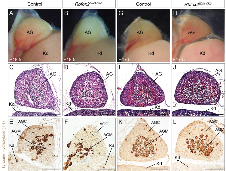
Figure 4—figure supplement 5. Neural crest contribution to adrenal gland is not affected in Rbfox2 knockout embryos.

Figure 4—figure supplement 5.

Morphological (A–B) and histological analyses (C–D) of the adrenal gland from E18.5 control (A, C and E) and Rbfox2 Pax3-CKO (B), (D), and F) embryos (n = 8 controls, n = 6 Rbfox2Pax3-CKO). Morphological (G–H) and histological analyses (I–J) of the adrenal gland from E17.5 control (G, I and K) and Rbfox2Wnt1-CKO (H), (J), and L) embryos (n = 4 controls, n = 4 Rbfox2Wnt1-CKO). Immunohistochemistry for tyrosine hydroxylase (E–F and K–L), a marker of neural crest-derived chromaffin cells, in sagittal sections of adrenal glands. Chromaffin cells of the adrenal medulla develop normally in the absence of Rbfox2 (F and L). AG, adrenal glands; Kd, kidney; AGC, adrenal gland cortex and AGM, adrenal gland medulla. Scale bars are 100 μm respectively.
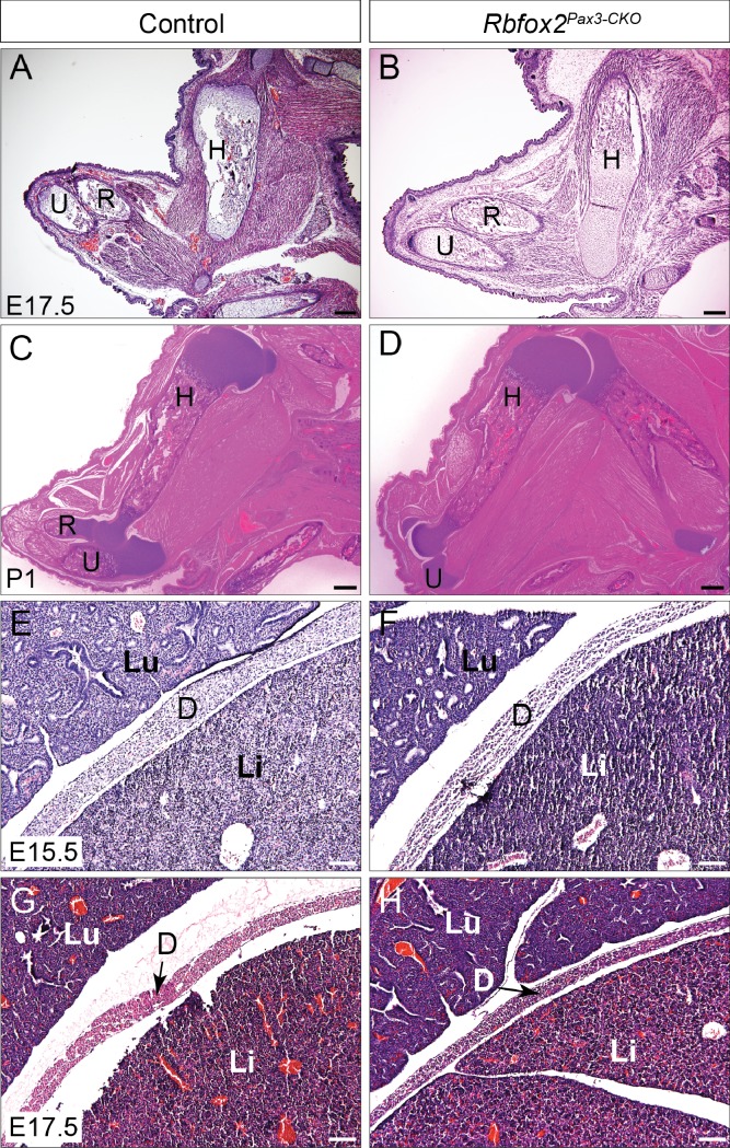
Figure 4—figure supplement 6. Limb and diaphragm musculature are not affected in Rbfox2Pax3-CKO embryos.

Figure 4—figure supplement 6.

Histological analysis of forelimb musculature from E17.5 control and Rbfox2Pax3-CKO embryos (n = 4 controls, n = 4 Rbfox2Pax3-CKO) (A–B). Histological analysis of forelimb musculature from P1 control and Rbfox2Pax3-CKO pups (n = 3 controls, n = 3 Rbfox2Pax3-CKO) (C–D). Histological analysis of diaphragm musculature from E15.5 (C and D) and E17.5 (E and F) control and Rbfox2Pax3-CKO embryos. H, Humerus; R, radius; U, Ulna; Lu, Lung; Li, Liver; D, Diaphragm. Scale bars are 200 μm (A–D) and 100 μm (E–H) respectively.