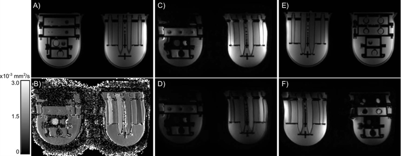
Figure 4:
Axial images from a central slice of the UCSF/NIST prototype breast phantom. The T1-weighted images (A, E) have no geometric distortion compared to the engineering designs, regardless of position in the coil. The EPI images (B-D, F) all demonstrate a geometric distortion in the right-left orientation: a stretch on the patient (and image) left and a shrinking on the patient (and image) right. In the ADC map (B), b-value = 0 image (C) and b-value = 800 s/mm2 image (D), the object on patient (image) left is larger than the object on patient (image) right. Similarly, when the objects are reversed (E, F), the object on patient (image) left is wider in the b-value = 0 s/mm2 image (F) compared to the T1-weighted image (E).

