. 2019 Jun 27;8:e46207. doi: 10.7554/eLife.46207

Figure 1. Mutations in helix-4 region of Snf7 induce cargo-sorting defect.

(A) Domain organization of Snf7, depicting the different helices (top). Bottom figure shows the sequence of helix-4, with the predicted helical motif highlighted with a box. Acidic residues are denoted in red, while cyan residues are basic amino acids. Bottom – structure of the helix-4 (from PDB 5fd7) in two orientations, highlighting the acidic residues on one surface. (B) Cartoon model of the polymeric arrangement of Snf7 in its linear form observed in the crystal lattice. (C) Canavanine sensitivity and Mup1-pHluorin flow-cytometry data (right) showing cargo-sorting/endocytosis defects of the helix-4 mutants of Snf7. Mup1-pHluorin data were collected 90 min after methionine addition. Error bars represent standard deviation from 3 to 7 independent experiments.

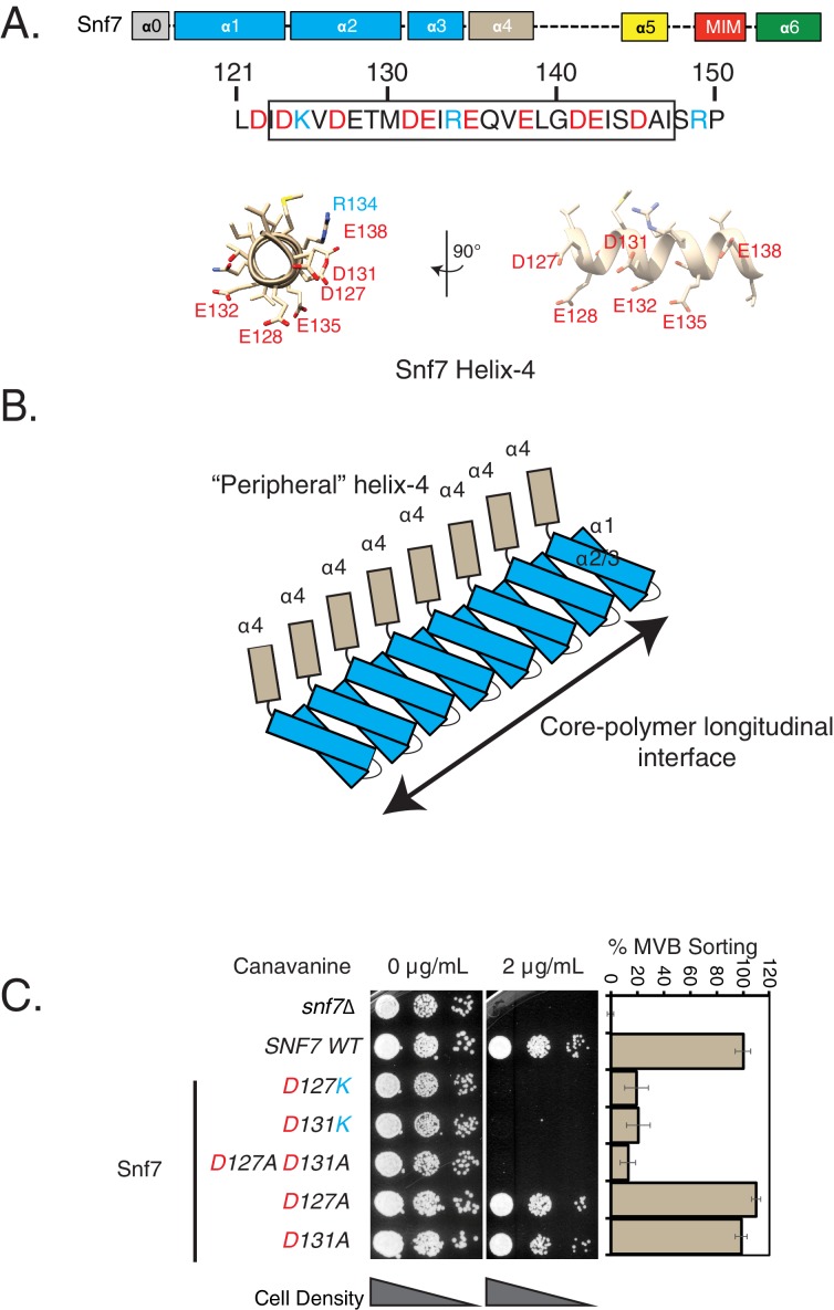
Figure 1—source data 1. Individual data points for data plotted in the figure (Figure 1C) for % MVB sorting.
DOI: 10.7554/eLife.46207.008

Figure 1.

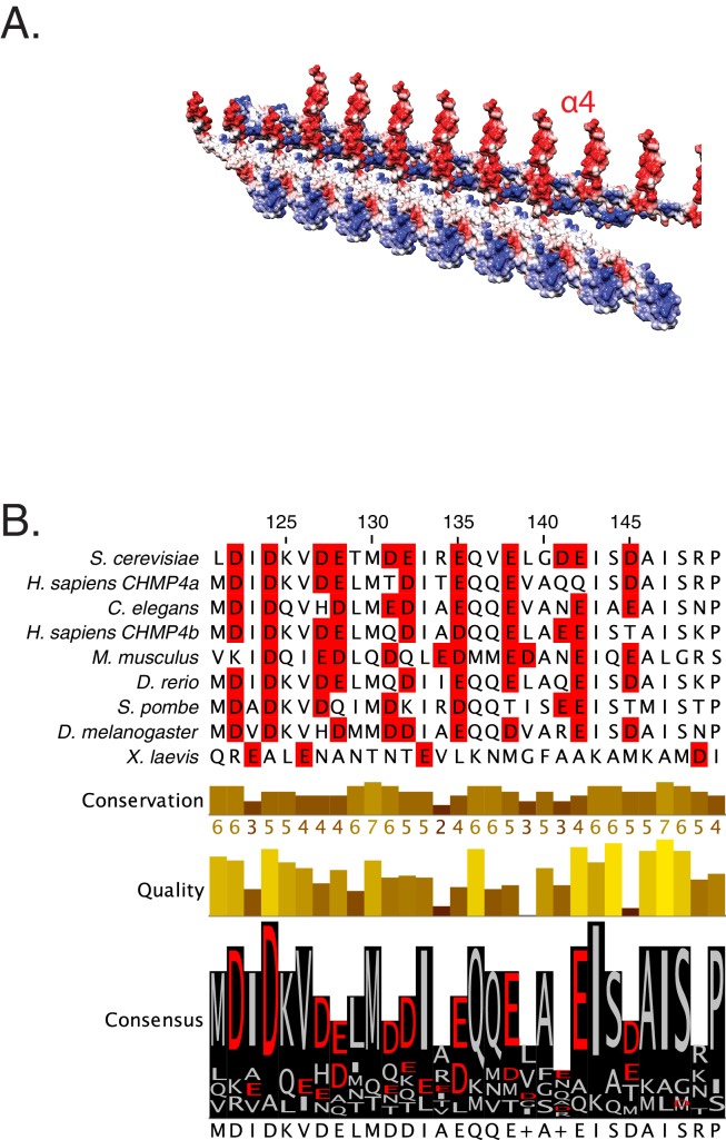
Figure 1—figure supplement 1. Snf helix-4 consists of conserved acidic residues.

Figure 1—figure supplement 1.

(A) Electrostatic depiction of the crystal lattice of Snf7 showing interaction between two laterally interacting strands of Snf7. Helix-4 (α4) is observed to be predominantly acidic in nature. (B) Sequence conservation of the helix-4 region of Snf7, with acidic residues highlighted in red.
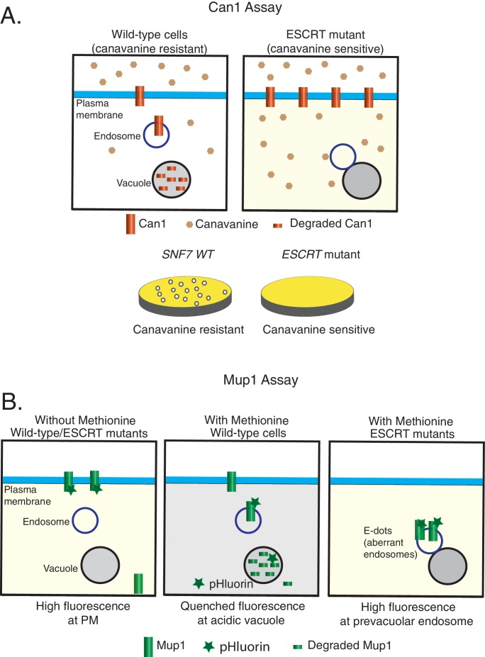
Figure 1—figure supplement 2. Models of the cargo-sorting assays used in this study using Saccharomyces cerevisiae.

Figure 1—figure supplement 2.

Canavanine-sensitivity assay (A) and Mup1-pHluorin assay (B). In ESCRT mutants, the model membrane proteins are unable to be trafficked to the vacuole for degradation.
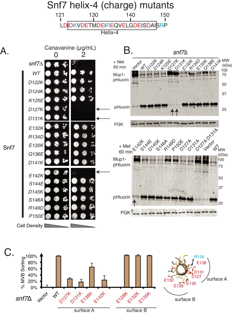
Figure 1—figure supplement 3. Mutational analysis of helix-4 residues of Snf7.

Figure 1—figure supplement 3.

Canavanine sensitivity assay (A) and Mup1-pHluorin degradation analysis (B) of several charge mutations in helix-4 residues of Snf7. Arrows point to the most sensitive mutations. (C) Mup1-pHluorin flow cytometry data of the mutations in two different surfaces (figure on the right) of Snf7 helix-4. Error bars represent standard deviation from three independent experiments.
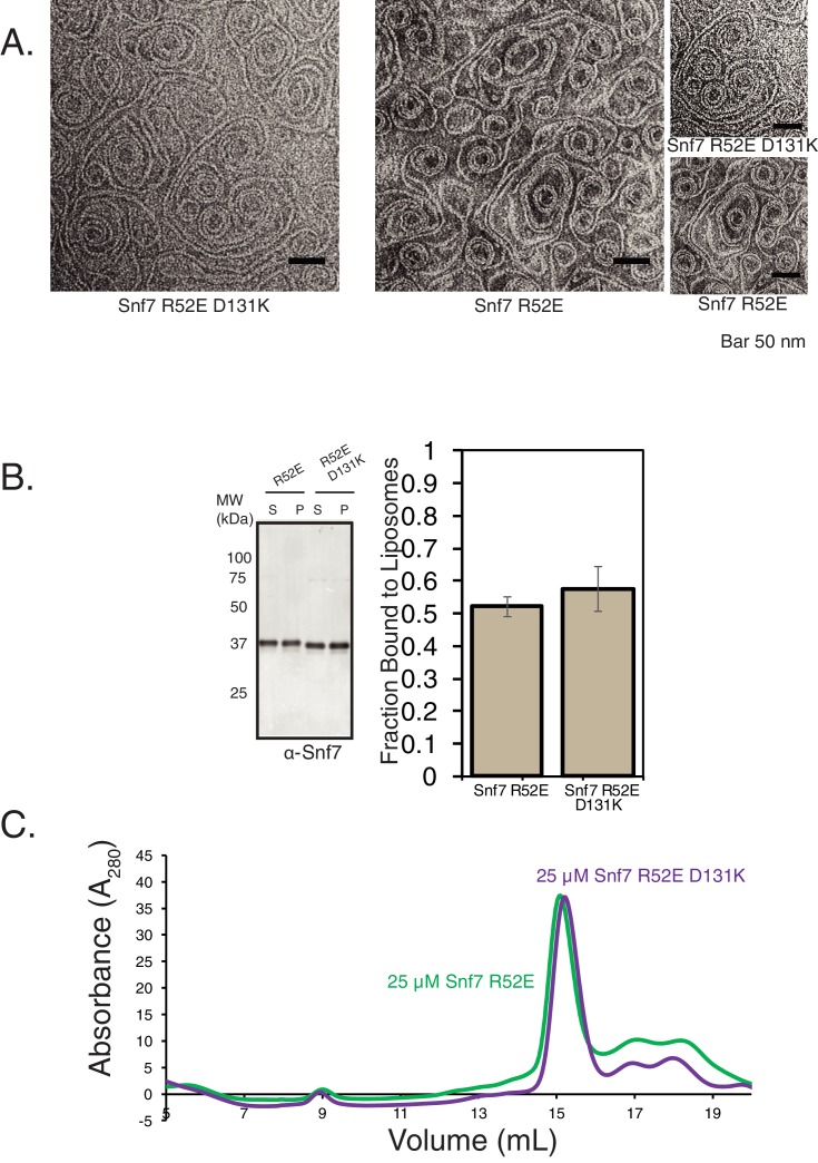
Figure 1—figure supplement 4. Helix-4 mutation does not affect stability of Snf7.

Figure 1—figure supplement 4.

(A) Electron microscopy (EM) images of Snf7 R52E and Snf7 R52E D131K mutations. Images on the far right are zoomed-in pictures. 10 μM Snf7 R52E and 10 μM Snf7 R52E D131K were used, incubated for 10 min on EM grids. (B) In vitro membrane-binding properties of Snf7 R52E and Snf7 R52E D131K. Experiments were performed with 200 nM of proteins and 0.5 mg/mL liposomes. Quantification on the right represent data from three technical replicates (three experiments with the same protein and lipid preparation samples). (C) Size-exclusion profiles of Snf7 R52E and Snf7 R52E D131K performed using an SD200increase column.