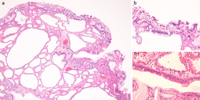
Figure 6.
Histopathological appearance of the lesion of Fig. 1. a: Hyperplastic change and cystic dilation of the fundic gland were observed [Hematoxylin and Eosin (H&E) staining, original magnification ×40]. b, c: An increase in the quantity of the chromatin, nuclear swelling, and front formation in a part of the foveola epithelium were recognized (H&E staining, original magnification b: ×100, c: ×200).

