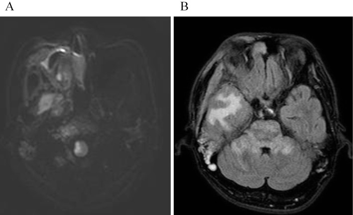
Figure 4.
(A) Diffusion-weighted image after the onset of lateral medullary syndrome (LMS) shows a hyperintense area in the right lateral medulla. (B) A few days after the onset of LMS, fluid attenuation inversion recovery image shows hyperintense areas in the right temporal lobe, the pons, and the bilateral middle cerebellar peduncles. The hyperintense white matter of the right temporal lobe is caused by the pressure of the extraosseous soft-tissue masses.

