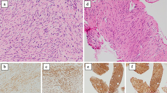
Figure 3.
Pathological findings of the tumors: (a-c) bladder tumor, (d-f) duodenal tumor, (a, d) Hematoxylin and Eosin staining ×400, (b, e) KIT, (c, f) CD34. Fascicular proliferation of spindle-shaped tumor cells was visible. No mitotic figures were observed (0/10 HPFs). Immunohistochemically, tumor cells stained positive for KIT, CD34, PDGFRA, DOG1, and vimentin, and stained negative for S-100 protein. The Ki-67 index of the tumor cells was 1%.

