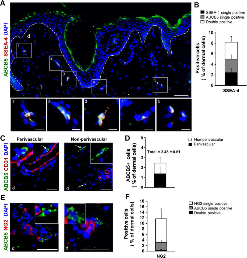
Figure 1. In situ characterization of ABCB5+ cells in their endogenous niche in healthy human skin.
(A-B) A microphotographic overview of healthy human skin subjected to immunostaining for ABCB5 (green) and the adult stem cell marker SSEA4 (red) revealed a distinct subpopulation of dermal cells positive for both markers (yellow overlay). Quantification of ABCB5 positive and SSEA-4 positive proved that 94.39% of dermal cells expressing ABCB5 were co-positive for SSEA-4. (C) Microphotographs of human skin subjected to immunostaining for ABCB5 (green) and the endothelial marker CD31 (red) revealed both a perivascular (left) and a dispersed interfollicular dermal localization (right) of ABCB5+ cells (white arrows), while no co-localization of ABCB5 and CD31 was observed. (D) ABCB5+ cells occurred at an average percentage of 2.45 0.61 of all dermal cells as determined in 8–10 microscopic fields of skin sections from 10 different donors. ABCB5+ cells were more abundant in a perivascular localization compared to a non-perivascular localization. (E) Double immunofluorescence staining for ABCB5 (green) and the pericyte marker NG2 (red) shows that perivascular ABCB5+ cells (white arrows) are distinct from NG2+ pericytes. (F) Quantification of ABCB5 positive and NG2 positive depict that only 0.32% of total dermal cells co-expressed ABCB5 and NG2 as determined in more than 20 microscopic fields of skin sections from 5 different donors. (A, B, D) Nuclei of all studied skin sections were counterstained with DAPI (blue). Scale bars: 50 μm; e = epidermis; d = dermis. Dashed white lines delineate epidermal from dermal layers.

