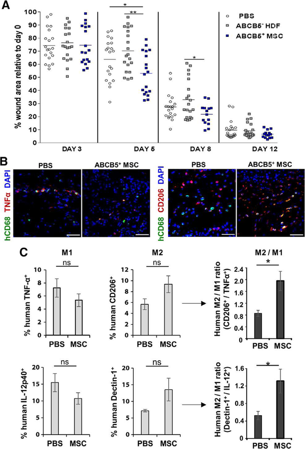
Figure 7. ABCB5+-derived MSCs stimulate a shift from human M1 macrophages to M2 macrophages in the humanized NSG model.
(A) Treatment of NSG mouse wounds with ABCB5+ MSCs led to a significantly reduced wound size compared to control groups at day 5 and day 8. (B) Representative microphotographs of day 5 wounds in humanized NSG mice show human CD68+ macrophages (green) in the wound bed with increased numbers of human inflammatory TNFα+ (red) macrophages in PBS treated wounds, compared to ABCB5+-derived MSC treatment (left panel). Co-immunostaining with M2 marker CD206 (red) displays an MSC-dependent increase in iron over-load wounds (right panel). Nuclei are counterstained with DAPI (blue). Scale bars = 50 μm. (C) Flow cytometry analysis of human CD68+-gated macrophages confirmed the ABCB5+ MSC-dependent phenotype shift from M1 to M2 with two different co-stainings. The ratios of both Dectin-1+/IL-12p40+ human macrophages and CD206+/TNFα+ increased after treatment with ABCB5+ MSCs. ns = not significant; * p < .05; ** p < .01, t-test.

