
Figure 2.
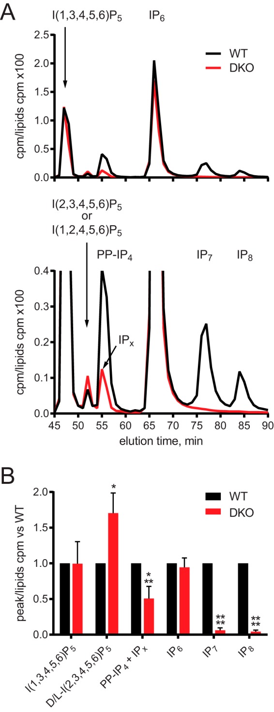
No change in IP6 or I(1,3,4,5,6)P5 levels in DKO cells. A, SAX-HPLC of myo-[3H]inositol–labeled cells. Cells were labeled for 5 days in inositol-free DMEM. Peaks of I(1,3,4,5,6)P5, D/l-I(2,3,4,5,6)P5 (the stereoisomers I(1,2,4,5,6)P5 and I(2,3,4,5,6)P5 cannot be distinguished by this HPLC method), 5PP-IP4, IP6, IP7, and IP8 were identified based on elution time compared with standards. The IP6K product 5PP-IP4, lost in DKO cells, coeluted with an unknown peak designated IPx. The data are shown fully (top) and zoomed-in to better visualize the smaller peaks (bottom). B, comparison of IPs species, normalized to WT to allow for differences in labeling between experiments. Bar chart shows mean ± S.D. from 3 or more experiments. HPLC trace in A is representative of an experiment performed 3 times. *, p < 0.05; ***, p < 0.001; ****, p < 0.0001, ANOVA with Tukey post test.