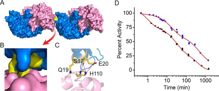
Figure 4.
Tandem dimers from ACMSD crystal structures. A, surface structures of two dimers of ACMSD (PDB entry 2HBV). Monomers in each dimer are colored blue and pink. B, zoom-in image of the dimer–dimer interface; the interacting area is highlighted in yellow. C, residues involved in the dimer–dimer interaction networks. D, comparison of the dissociation profiles of wtACMSD (black squares) and the mutant H110A (blue squares) as measured by specific activity.

