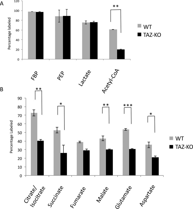
Figure 1.
Flux of [U-13C]glucose in TAZ-KO cells relative to WT. A and B, the percentage of 13C-labeled metabolites was measured after 1-h incubation with [U-13C]glucose. Metabolites from glycolysis (A) and the TCA cycle (B) were analyzed by LC-MS and GC-MS. Data shown are mean ± S.D. (n = 3). *, p < 0.05; **, p < 0.01; ***, p < 0.001.

