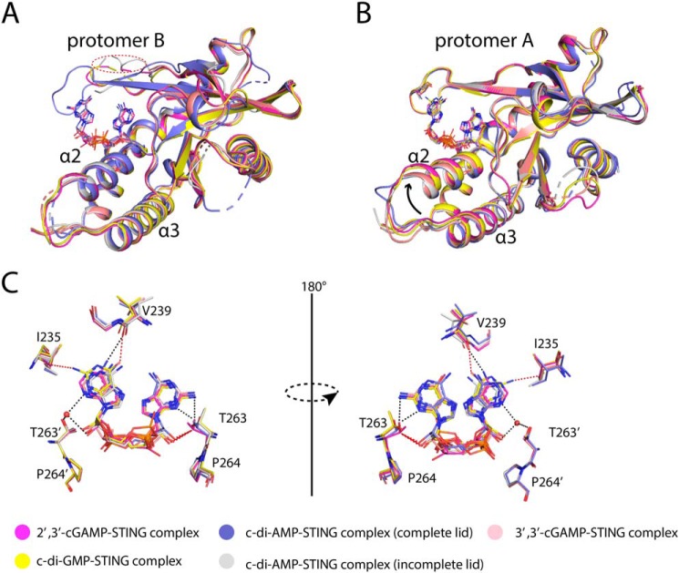
Figure 11.
Structural comparison of porcine STINGCBD–CDNs complex. A, structural comparison of protomer B of porcine STINGCBD–CDNs complex. The lid regions of protomer B are incomplete except one of porcine STINGCBD–c-di-AMP complex. The red dashed circle shows the disordered lid region in protomer B. B, structural comparison of protomer A of porcine STINGCBD–CDN complex. The tilt of α2–α3 in 2′,3′-cGAMP is the most dramatic one. The black arrow indicates the upward movement of α2–α3. C, superimposition of CDNs shows the 2′,3′-cGAMP is the favorite ligand for STING. The black dashed line represents the hydrogen bond. The red dashed line represents the repulsive van der Waals contact (bad contact or bump).

