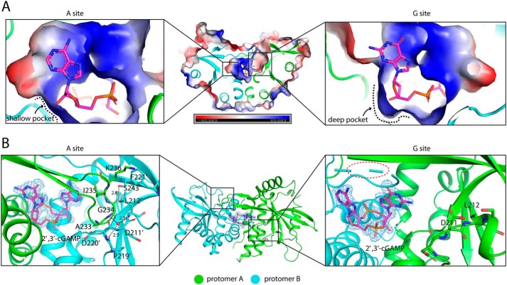
Figure 4.
Asymmetric ligand-binding pocket in porcine STINGCBD. Protomer A and B are colored green and cyan, respectively. 2′,3′-cGAMP is shown as a magenta stick. A, cross-section of 2′,3′-cGAMP–binding pocket in porcine STINGCBD. Left panel shows the complete A site, and right panel shows the incomplete G site. Electrostatic potential is shown. B, left panel shows the interactions between hairpin tip of protomer A and protomer B. The water molecule is shown as a red sphere. The black dashed lines represent the hydrogen bond, and the values show the length of hydrogen bond with the unit of Å. The simulated annealing omit Fo − Fc electron-density map for 2′,3′-cGAMP (blue mesh) is contoured at 3 σ. Middle panel shows the top view of porcine STING-2′,3′–cGAMP complex. Right panel shows the interaction between disordered hairpin of protomer B and protomer A.

