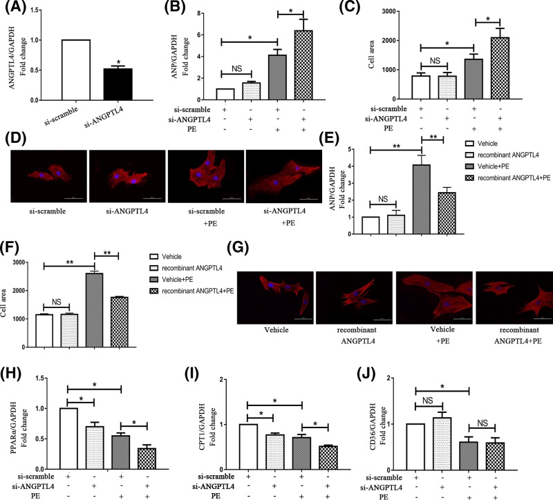
Figure 2. Effects of ANGPTL4 knockdown and recombinant ANGPTL4 treatment on PE-induced cardiomyocyte hypertrophy and the genes (PPARα, CPT-1 and CD36) involved in the regulation of FAO.
Cardiomyocytes were transfected with siRNA and incubated with or without PE for 24 h in serum-free medium. (A) The siRNA-mediated knockdown of ANGPTL4 was confirmed by q-PCR. (B) The effect of siRNA-mediated knockdown of ANGPTL4 on ANP mRNA expression was determined by q-PCR, and GAPDH was used as an internal control. (C) The effect of silencing ANGPTL4 on the cell surface area. After siRNA transfection, cardiomyocytes were treated with or without PE for 24 h. (D and G) Cardiomyocytes were stained with troponin I, and the nuclei were stained with DAPI. (E) The effect of recombinant ANGPTL4 on ANP mRNA expression was detected by q-PCR. (F) The effect of recombinant ANGPTL4 on the cell surface area. After siRNA transfection, the expression levels of PPARα (H), CPT-1 (I) and CD36 (J) were detected by q-PCR, and GAPDH was used as an internal control. *P<0.05 versus the corresponding control group; NS indicates no significance versus the corresponding control group. Each of the experiments was repeated four to seven times; n = 4–7.

