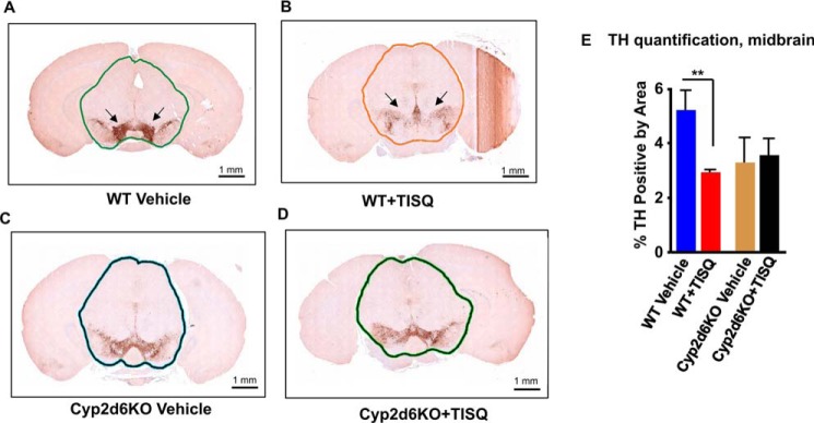
Figure 4.
The role of CYP2D6 in TISQ-induced brain damage and neuronal loss. WT (n = 6) and Cyp2d6KO (n = 6) mice were injected i.p. with TISQ (64 mg/kg b.w., n = 3) or vehicle (n = 3) once a day for 21 days. Brains were extracted following euthanasia, and formalin-fixed brains were sliced using the coronal brain matrix system as described under “Experimental procedures.” The brain slices were stained with TH antibody as described under “Experimental procedures.” IHC evaluation was performed on two slides per sample, two serial sections per slide, with an ∼20-μm step between slides. A and C, WT and Cyp2d6KO mouse brains stained with TH antibody. B and D, TISQ-treated mouse brains. E, % TH positive by area, mean ± S.D. of TH-positive neurons. **, p < 0.01 versus vehicle. n represents the number of mice used in each group.

