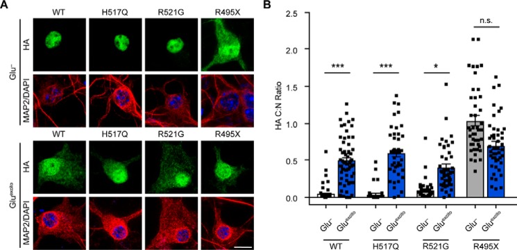
Figure 3.
Effect of Gluexcito on ALS-linked FUS variants. A, cortical neurons transfected with the indicated FLAG–HA-tagged FUS variants were exposed to Gluexcito, and nuclear FLAG–HA–FUS egress was assessed by immunofluorescence. Exogenous FUS was detected using an anti-HA antibody (green) within MAP2-positive neurons (red). Nuclei were stained with DAPI (blue). Scale bar = 10 μm. B, quantification of the C:N ratio for FLAG–HA–FUS variants in A revealed a significant shift in equilibrium toward the cytoplasm for FLAG–HA–FUS WT, H517Q, and R521G, but not R495X (Student's t test; WT, ***, p = 0.0003; H517Q, ***, p = 0.0003; R521G, *, p = 0.0197, n.s. = not significant, n = 3–5 biological experiments). However, the C:N ratio of H517Q, R521G, and R495X was not significantly different from FLAG–HA–FUS WT following Gluexcito treatment (Glu−; one-way ANOVA and Dunnett's post hoc test; n.s. = nonsignificant, n = 3–5 biological replicates). Black squares represent individual, cellular C:N measurements. Error bars represent S.E.

