Figure 5.
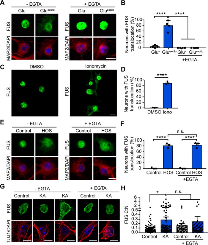
Calcium is necessary and sufficient for FUS translocation in primary cortical and motor neurons. A, reducing extracellular calcium levels with 2 mm EGTA attenuates FUS egress (green) in MAP2-positive neurons (red) following excitotoxic insult. Nuclei were stained with DAPI (blue). B, quantification of confocal microscopy findings in A confirmed the effect of EGTA treatment (two-way ANOVA and Tukey's post hoc test; for all statistical comparisons, ****, p < 0.0001; n = 4 biological replicates). C and D, application of 10 μm of the calcium ionophore, ionomycin (Iono), for 1 h induced FUS translocation relative to the DMSO control (Student's t test; ****, p < 0.0001; n = 3 biological replicates). E and F, FUS translocation induced by hyperosmotic stress (HOS) was not significantly attenuated by EGTA treatment (two-way ANOVA and Tukey's post hoc test; for all significant statistical comparisons, ****, p < 0.0001, n.s. = nonsignificant; n = 3 biological replicates). G and H, 10-min treatment of 300 μm kainic acid (KA) followed by a 1-h recovery induced FUS egress in primary motor neurons. Neurons were identified using the neuronal marker, TUJ1 (red), and nuclei were stained with DAPI (blue). H, kainic acid–induced FUS egress was statistically significant relative to the washout control; however, the addition of EGTA did not significantly restore FUS localization (two-way ANOVA and Tukey's post hoc test; *, p < 0.0190, n.s. = nonsignificant; control and KA, n = 8 biological replicates, EGTA and KA + EGTA n = 3 biological replicates). Black squares indicate individual cell measurements normalized to the average of the replicate control. Accordingly, means represent the normalized average of biological replicates. Error bars represent S.E. Scale bars = 10 μm.

