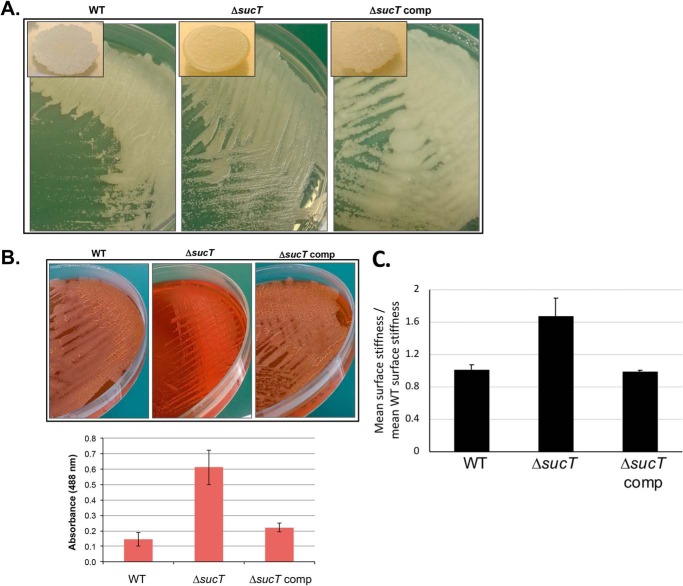
Figure 5.
Alterations in the cell surface properties of the M. smegmatis sucT mutant. A, colony morphology of WT mc2155, mc2ΔsucT, and mc2ΔsucT/pMVGH1-sucT (ΔsucT comp) after 3 days of incubation on 7H11-OADC agar at 37 °C. B, Congo red binding on a TS agar plate and in TS liquid medium (graph). Shown on the graph are the average ± S.D. absorbances of acetone extracts measured for three biological replicates. C, analysis of the cell surface rigidity of the WT, mutant, and complemented mutant strains by correlated optical fluorescence and AFM. Two-sided rank sum test demonstrates a statistically significant difference in stiffness between the sucT mutant and both the WT (p = 0.0214) and the sucT complemented mutant (p = 0.0011).

