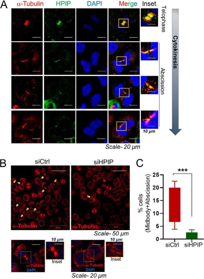
Figure 10.

HPIP localizes to midbody during cytokinesis and thus loss of HPIP expression leads to defects in cytokinesis. A, confocal images representing the localization of indicated proteins at telophase and cytokinesis in HeLa cells. Green, HPIP; red, α-tubulin; blue, DNA (DAPI) (magnification, 60×). Scale bar, 20 μm. B, confocal images representing the localization of tubulin in siCtrl or siHPIP transfected HeLa cells. Upper panels, magnification, 20×. Lower panels, magnification, 60×). C, data represent percentages of cells having midbody. ***, p < 0.001 was considered significant.
