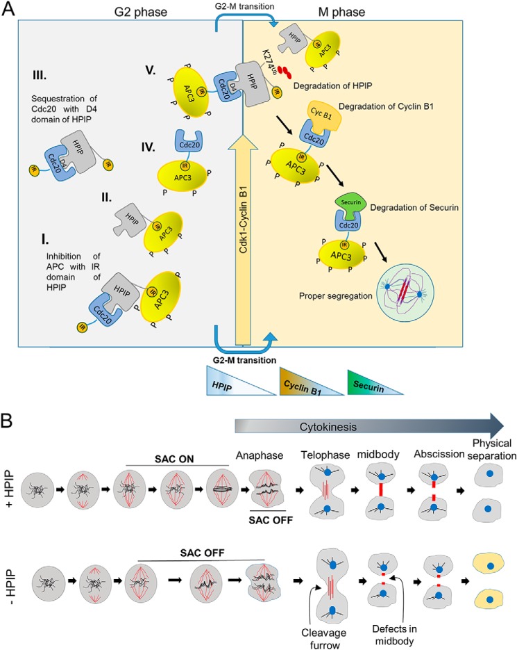
Figure 11.
Model represents the role of HPIP during cell cycle progression. A, schematic illustration showing the mechanistic role of HPIP in G2/M transition during mitosis. I, II, II, IV, and V are the possible protein complexes formed in a combinatorial fashion involving HPIP, APC, and Cdc20. HPIP promotes G2/M transition by stabilizing cyclin B1 during late G2 phase and itself is subjected to degradation during mitosis. Degradation of cyclin B1 and then Securin follows soon after the HPIP destruction by APC/Cdc20. D4 and IR motifs of HPIP function differently, because the D4 domain is primarily involved in HPIP stability, whereas the IR domain participates in inhibition of APC–Cdc20 activity. B, schematic illustration showing the role of HPIP in spindle checkpoint function and cytokinesis. In the presence of HPIP, the SAC is on, and cells proceed to cytokinesis with proper midbody/abscission and generate daughter cells with normal phenotype. In contrast, loss of HPIP results in SAC being off, defects in midbody formation, and thus abnormal cells.

