Figure 3.
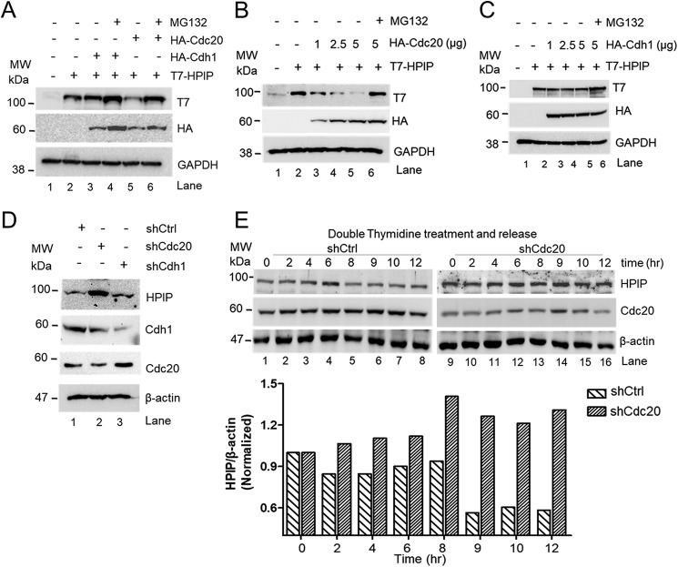
Cdc20 but not Cdh1 stimulates HPIP degradation. A, HEK293T cells were co-transfected with T7–HPIP and HA–Cdc20 or HA–Cdh1 plasmid constructs. Following MG132 (10 μm) treatment, cell lysates were blotted as indicated. B and C, HEK293T cells were co-transfected with T7–HPIP and increasing concentrations of either HA–Cdc20 (B) or HA–Cdh1 (C) (1–5 μg) plasmid constructs. After 8 h of MG132 treatment, cell lysates were blotted as indicated. D, HeLa cells were transfected with control shRNA, Cdc20 shRNA, or Cdh1shRNA, and cell lysates were analyzed by Western blotting as indicated. E, Cdc20 shRNA-treated cells were synchronized and released at indicated time points and blotted as indicated (upper panel). The bar graph showing the quantification of HPIP protein band intensity (lower panel) (n = 2). Ctrl, control; GAPDH, glyceraldehyde-3-phosphate dehydrogenase; sh, short hairpin; MW, molecular weight.

