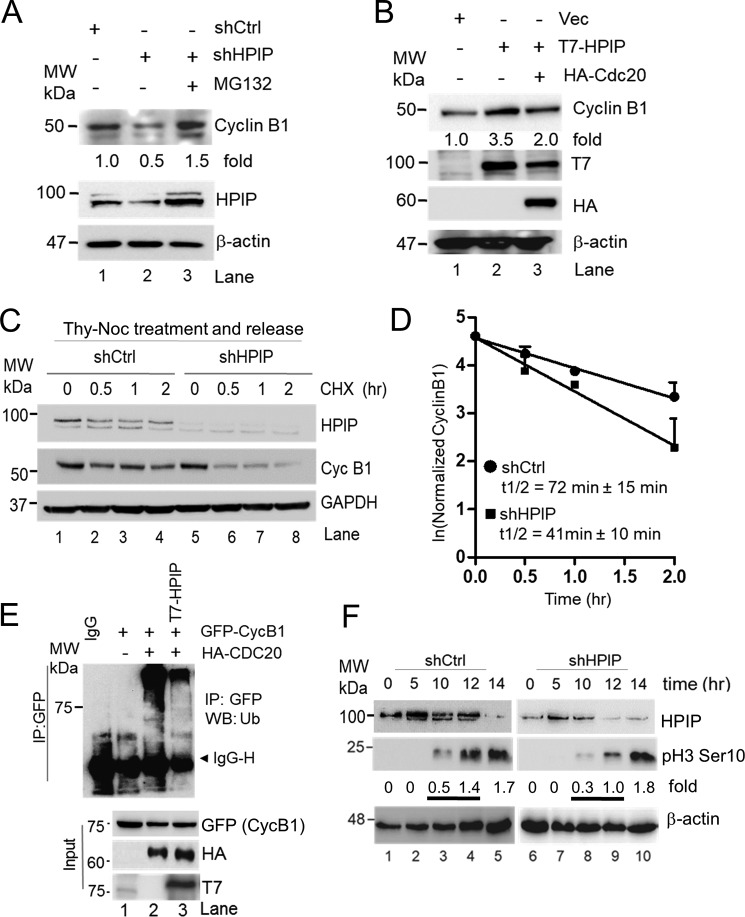
Figure 6.
HPIP stabilizes cyclin B1 during G2/M transition. A, HeLa cells transfected with shCtrl (lane 1) and shHPIP (lanes 2 and 3) were treated with MG132 (lane 3) and blotted as indicated. B, HEK293T cells were transfected with T7–HPIP alone or in combination of T7–HPIP and HA–Cdc20, and cell lysates were blotted as indicated. C, stably knocked down HeLa cells with indicated shRNAs were synchronized by thymidine–nocodazole block and treated with cycloheximide (20 μg/ml) for the indicated time points. Cell lysates were then analyzed by Western blotting. D, the line graph represents the quantification of cyclin B1 protein bands from C. E, HEK293T cells were co-transfected with GFP–cyclin B1 or HA–Cdc20 and with or without T7–HPIP. 48 h post-transfection, cell lysates were T7-immunoprecipitated and blotted as indicated. F, double thymidine block synchronized HPIP-depleted HeLa cells were released in presence of nocodazole (100 ng/ml) at the indicated time points and analyzed by Western blotting as indicated. Ctrl, control; GAPDH, glyceraldehyde-3-phosphate dehydrogenase; IP, immunoprecipitation; sh, short hairpin; MW, molecular weight; Vec, vector.

