Figure 7.
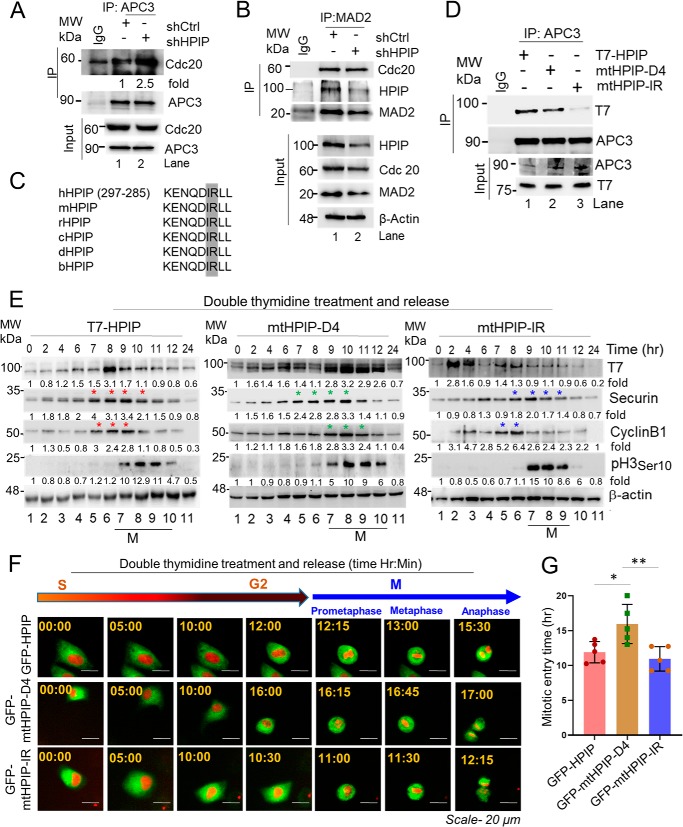
HPIP binds and inhibits APC/C–Cdc20 activity through IR motif. A and B, HeLa cells stably transfected with either shCtrl or shHPIP were lysed, and cell lysates were subjected to co-IP by APC3 (A) or MAD2 (B) followed by Western blotting as indicated. C, conservation of IR motif in HPIP among various species. D, HeLa cells transfected with wtHPIP, mtHPIP–D4, or mtHPIP–IR were subjected to co-IP with APC3 antibody followed by Western blotting as indicated. E, HeLa cells transfected with wtHPIP, mtHPIP–D4, or mtHPIP–IR were subjected to double thymidine synchronization followed by release at the indicated time points and blotted as indicated. An asterisk denotes expression of the indicated proteins at peak in the specified time period. F, representative time-lapse live cell fluorescent images of GFP–HPIP, GFP–mtHPIP–D4, or GFP–mtHPIP–IR transfected HeLa–H2B–mCherry (red) cells that are synchronized by DT block at the S phase followed by release into fresh medium and captured at indicated time points (magnification, 20×). G, quantification data of F. The quantified results are presented as means ± S.D. using Student's t test. *, p < 0.01; **, p < 0.001 were considered significant. Ctrl, control; IP, immunoprecipitation; MW, molecular weight; sh, small hairpin.

