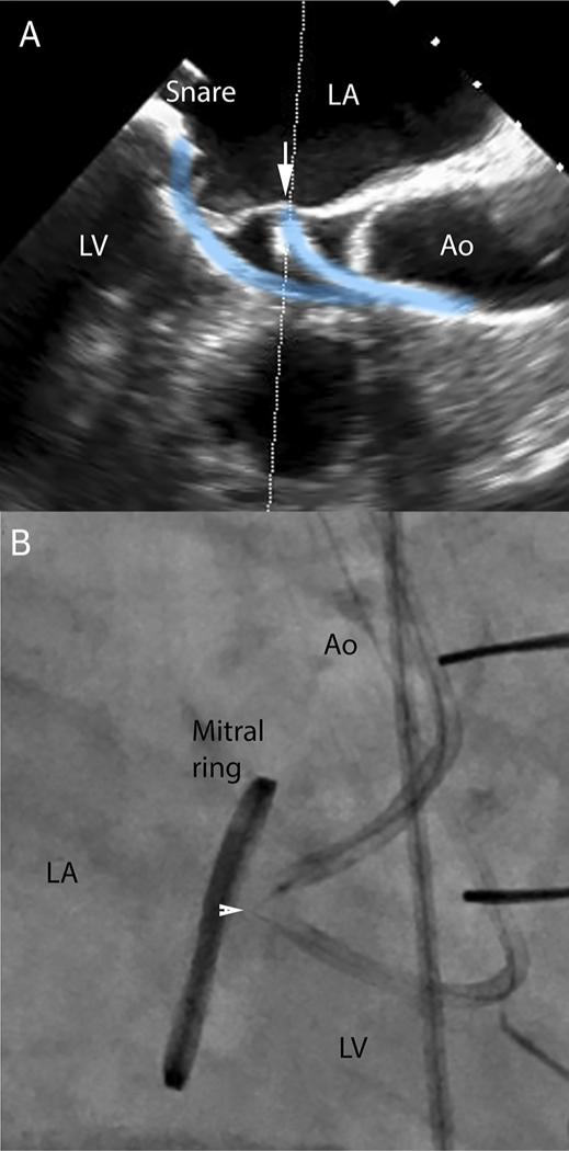
FIGURE 2. LAMPOON Procedure.

(A) Transesophageal echocardiography visualization of retro-grade LVOT catheter contacting the middle of the anterior mitral leaflet (arrow) and pointed toward a snare positioned by a second retrograde catheter in the left atrium. (B) The focally denuded lacerating guidewire straddles the anterior mitral leaflet (arrowhead) during electrification to achieve laceration (Online Video 1). Ao = Aorta; LA = left atrium; LV = left ventricle.
