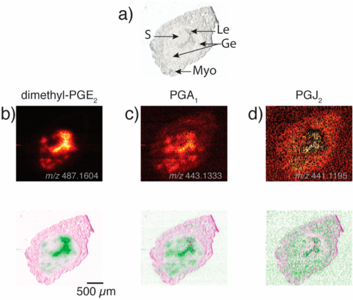
Figure 4.
Ion images for tentatively assigned PG species in Wnt5ad/d mouse uterine tissue on day 4 of pregnancy, (a) optical image, (b, c, and d) ion images and false color fused image for dimethyl-PGE2 (m/z 487.1604, [C22H36O5Ag]), PGA1 (m/z 443.1333, [C20H30O4Ag]+), and PGJ2 (m/z 441.1195, [C20 H32O4Ag]+), respectively. All ion images are for 109Ag adducts and have been normalized to the internal standard PGE2-d9 as discussed in the Supporting Information and Figure S9. Brighter colors correspond to higher signal intensities. Le, luminal epithelium; Ge, glandular epithelium; S, stroma; and Myo, myometrium.46

