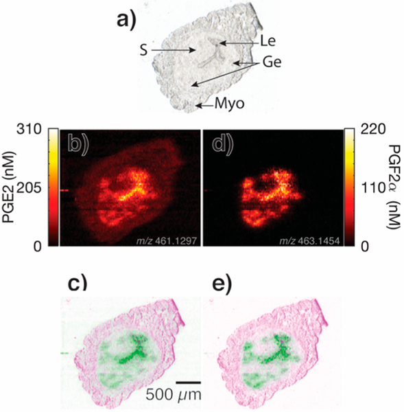
Figure 5.
Quantitative ion images for PGE2 and PGF2α from Wnt5ad/d mouse uterine tissue on day 4 of pregnancy, (a) optical image, (b) ion image for PGE2 (109Ag+ adduct), and (c) resulting false color fused image detailing PGE2 localization. The ion image and false color image for PGF2α are shown in (d and e, respectively). Le, luminal epithelium;Ge, glandular epithelium; S, stroma; and Myo, myometrium.46

