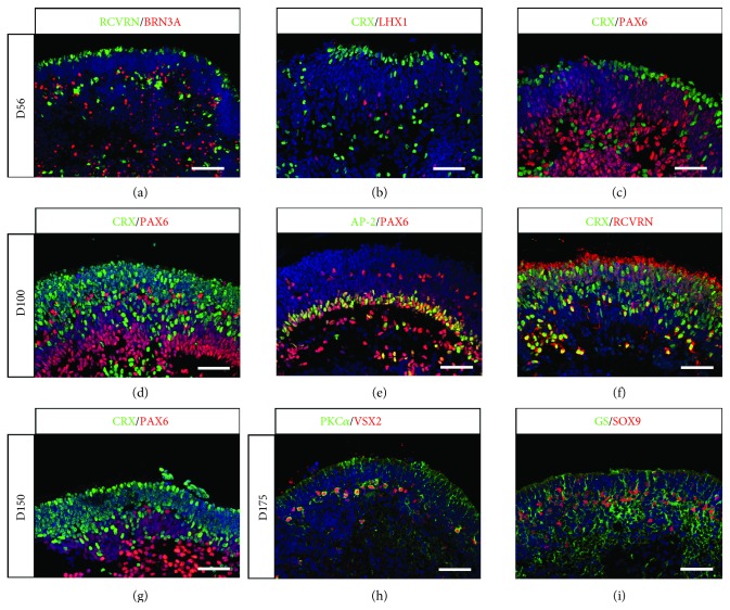
Figure 3.
Generation of pseudolaminated retinal organoids containing all retinal cell types from human MGC-derived iPSCs. (a-f) Immunofluorescence staining of cryosections from retinal organoids at D56 (a-c) and D100 (d-f) using markers for retinal ganglion cells (BRN3A, PAX6), horizontal cells (LHX1, PAX6), amacrine cells (AP2, PAX6), and photoreceptors (CRX, RCVRN). (g-i) Immunofluorescence staining of cryosections from retinal organoids at D150 (g) and D175 (h, i) using markers for photoreceptors (CRX), bipolar cells (VSX2 and PKCα), and MGCs (GS and SOX9). Nuclei were counterstained with DAPI (blue) (scale bars: a, 100 μm; b-i, 50 μm).

