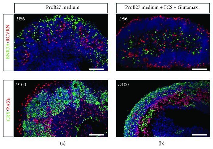
Figure 4.
Improvement of retinal organoid lamination in the presence of FCS and Glutamax. Immunofluorescence staining of cryosectioned retinal organoids from human MGC-derived iPSCs using markers for retinal ganglion cells (BRN3A, PAX6), amacrine/horizontal cells (PAX6), and photoreceptors (CRX) after 56 and 100 days in floating cultures in the absence (a) or in the presence (b) of 10%FCS and 2 mM Glutamax in the ProB27 medium. Nuclei were counterstained with DAPI (blue) (scale bars: 100 μm).

