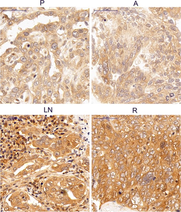
Fig. 1.

IHC staining for the LPAR1 protein in four types of matched lesions from a patient with OSC. In the image of IHC staining from the same patient, increased LPAR1 staining was observed in the recurrent lesions and lymphatic metastatic lesions compared with the primary tumor lesions. However, no differences in LPAR1 expression were observed between the primary tumor lesions and abdominal disseminated lesions. Original magnification: ×400. P primary tumor samples, A abdominal disseminated samples, LN lymph node metastases samples, R recurrent samples
