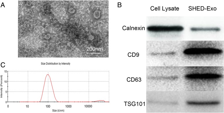
Fig. 2.
Identification of SHED-Exos. a Morphology of the exosomes according to TEM. The exosome vesicles were with diameters ranging from 30 to 100 nm. Scale bars indicate 200 nm. b Surface exosomal markers of SHED-Exos (CD63, TSG101, and CD9) were quantified by western blotting. c Determination of the concentration and particle size of SHED-Exos using a Nanosight Tracking Analysis system

