Fig. 1.
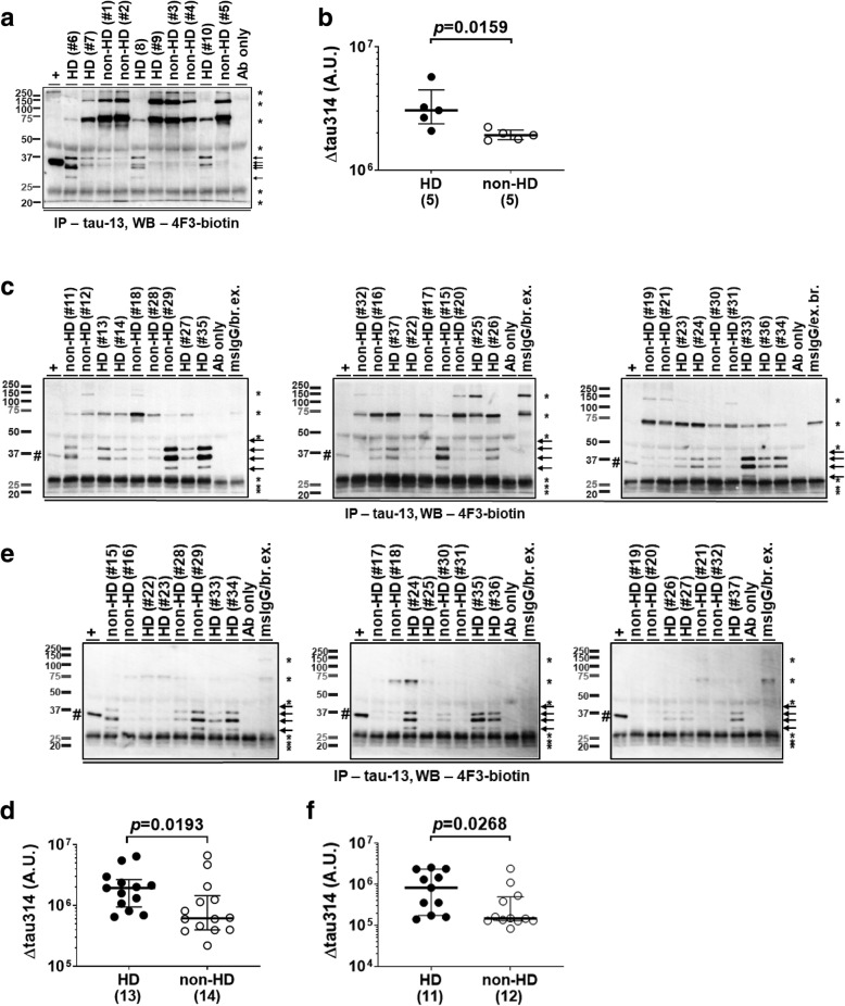
Levels of Δtau314 proteins are elevated in HD patients. a, c, e Representative IP/WB showing that the tau-13-immunoprecipitated Δtau314 proteins (arrows) were detected by the biotin-conjugated 4F3 antibody in the prefrontal cortex (Brodmann’s area (BA) 8) of subjects from the HUB-ICO-IDIBELL Biobank, Spain (a), in the prefrontal cortex (BA 8/9) of subjects from the NIH NeuroBioBank and the New York Brain Bank (c), and in the caudate nucleus of subjects from the NIH NeuroBioBank (e). HD, Huntington’s disease patients; non-HD, individuals without Huntington’s disease. +, in vitro synthetic Δtau314 proteins (hash; 20 μL of sample used in IP, positive control). *, non-specific bands. Sample IDs and disease diagnoses (Additional file 1: Table S1) were shown for IP reactions. Ab only, IP reaction containing only 10 μg of tau-13 (negative control). MsIgG/br. ex., IP reaction containing 10 μg of mouse IgG and 173 μg of soluble brain proteins (negative control). For the MsIgG/br. ex. lanes in figure c, brain extracts were a mixture of the nine samples studied in the same blot with each contributing 1/9th the amount (i.e., 19.2 μg); for the MsIgG/br. ex. lanes in figure e, 1/8th the amount (i.e., 21.6 μg). b, d, f Comparison of levels of Δtau314 proteins in the prefrontal cortex (BA 8) of HD patients and non-HD individuals from the HUB-ICO-IDIBELL Biobank, Spain (b), in the prefrontal cortex (BA 8/9), from the NIH NeuroBioBank and the New York Brain Bank (d), and in the caudate nucleus, from the NIH NeuroBioBank (f). The numbers of analyzed subjects are shown in parentheses. A.U. = arbitrary unit. The y-axes in figures are in log scale. Mann-Whitney test was used for between-group comparison; medians (middle long bars), and 1st (lower short bars) and 3rd (upper short bars) quartiles are shown

