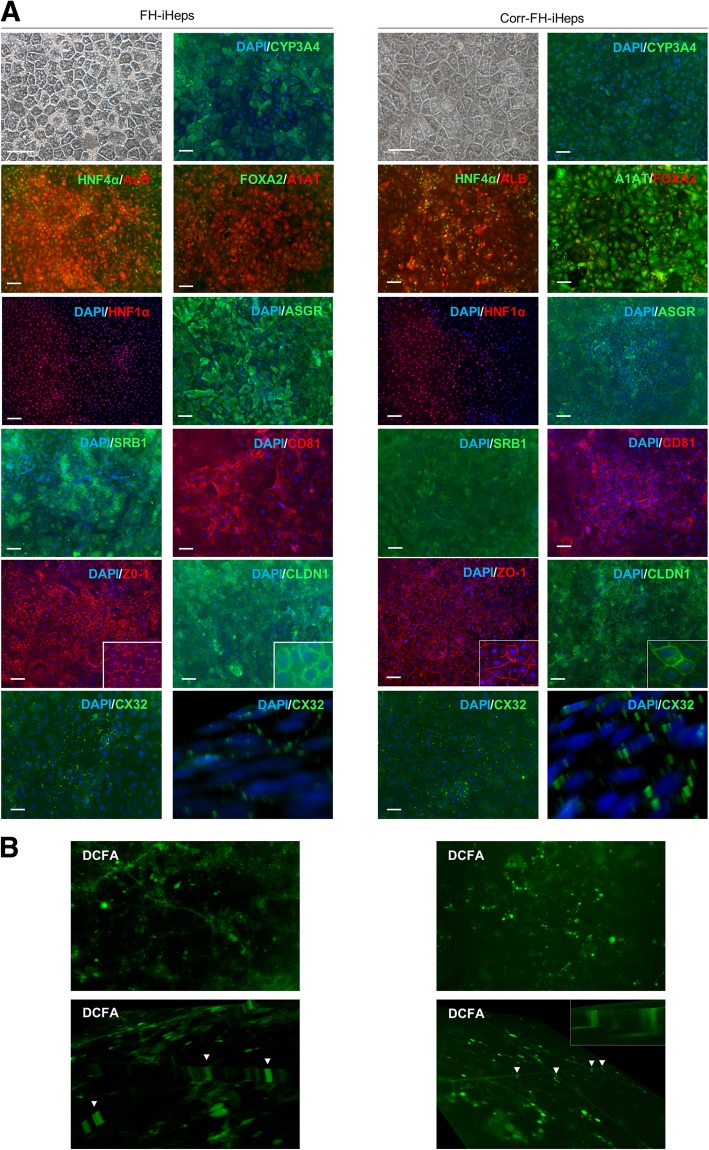
Fig. 3.
Differentiation of FH- and corr-FH-iPSCs into hepatocytes. a Representative pictures of cell morphology and immunostainings of the indicated markers at day 25 of differentiation. Scale bars: 50 μm. b Representative images of DCFA excretion at the biliary poles of corr-FH-iHeps. All images were taken with × 10 objective, z-stacks of xy sections of the cells were acquired with an epifluorescence microscope (Nikon Elipse) and analyzed with ImageJ software. Arrowheads indicate bile canaliculi

