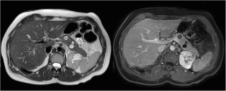
Fig. 2.

Abdominal stage. Transversal T2 image (left side) and T1 image with fat suppression after intravenous Gd administration in the portal-venous phase. The pancreas contains two small cystadenomas (11 and 13 mm) and a small, simple cyst (4 mm). The upper pole of the left kidney is distorted due to an irregularly enhancing mass (renal cell carcinoma)
